


Your muscle does something remarkable during an eccentric contraction: it produces more force while getting longer. That seems counterintuitive. Most people assume a contracting muscle is a shortening muscle. But the relationship between force and length is more nuanced than that, and understanding it is what separates surface-level knowledge from clinical depth.
Isotonic exercise sits at the heart of physiotherapy rehabilitation. Every squat, every bicep curl, every step up a staircase involves isotonic contractions: dynamic movements where a muscle generates tension and moves a joint through its full range. The word isotonic comes from the Greek for “same tension,” though in practice the load stays constant, not the tension itself. Both contraction types have distinct physiological mechanisms and distinct clinical applications, and knowing the difference shapes how exercise is prescribed at every stage of recovery.
What Is Isotonic Exercise?
Isotonic exercise is a form of dynamic resistance exercise in which a muscle generates force while changing length against a constant external load. It includes two contraction types: concentric, where the muscle shortens as it overcomes resistance, and eccentric, where it lengthens under load. According to the American College of Sports Medicine, both types produce meaningful adaptations in muscular strength, hypertrophy, and endurance, making isotonic exercise the foundational modality in physiotherapy rehabilitation.
Isotonic exercise differs from the other major contraction types used in clinical practice. Isometric exercise generates force with no change in muscle length and no joint movement: a static wall squat is isometric. Isokinetic exercise produces movement at a constant velocity, typically via specialised dynamometer equipment, and is used primarily in clinical assessment rather than routine rehabilitation. Isotonic exercise occupies the middle ground: the load is fixed, velocity is free to vary, and the muscle works dynamically through its full functional range.
For students new to exercise prescription, the most practical clinical understanding is this: if a movement involves a joint changing angle against resistance, it is isotonic. That covers the vast majority of rehabilitation exercises performed outside a testing laboratory.
According to the American College of Sports Medicine’s 2009 position stand on resistance training, progressive isotonic exercise produces measurable adaptations in muscular strength, power, hypertrophy, and endurance, making it the foundational training modality for rehabilitation, athletic conditioning, and general physical health.
What Actually Happens Inside the Muscle During Isotonic Exercise?
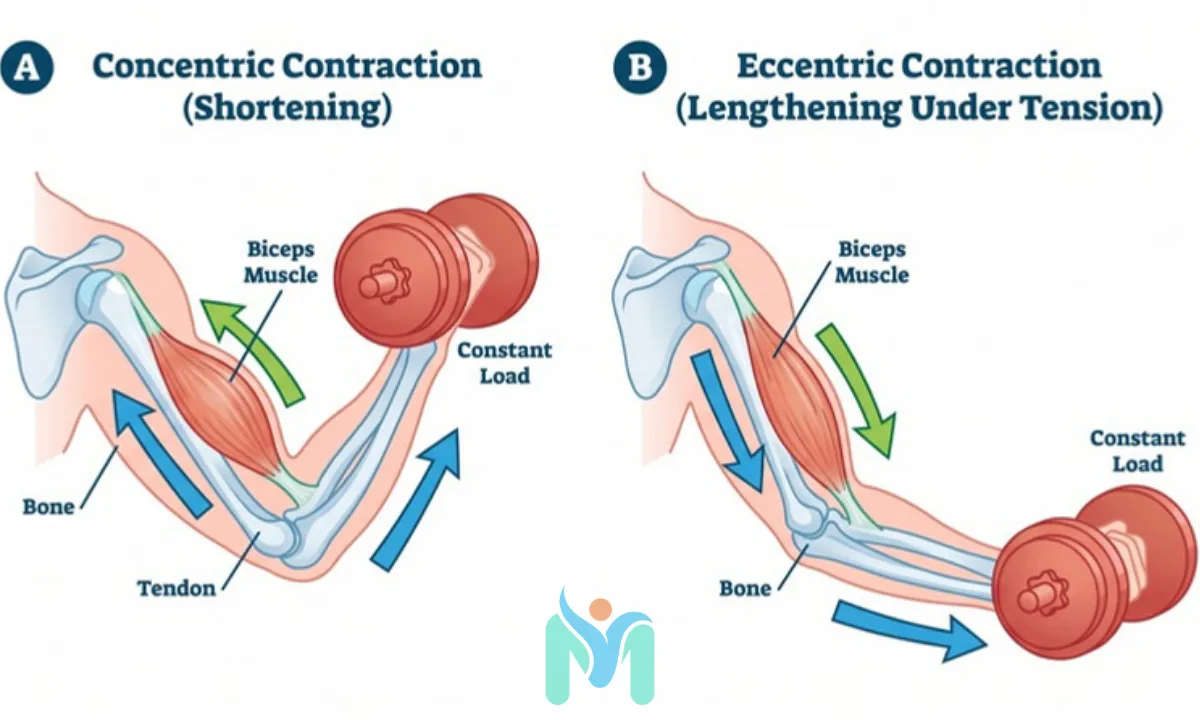
During a concentric contraction, the sarcomere (the basic contractile unit of muscle tissue) shortens as actin and myosin filaments slide together. The muscle generates enough force to exceed the external load, and the joint moves in the direction of the contraction. Think of the upward phase of a bicep curl: the elbow flexors shorten, the forearm rises, the resistance is overcome.
During an eccentric contraction, the sarcomeres lengthen while still under tension. The muscle does not switch off: it resists the load in a controlled way, like the lowering phase of the same bicep curl. Cross-bridges between actin and myosin are forcibly extended, and the passive elastic structures within the muscle, particularly the giant structural protein titin, contribute additional force. Research suggests that titin functions as a molecular spring during eccentric loading, storing elastic energy in a way that is not possible during concentric work, according to a 2014 review by Herzog in the Journal of Experimental Biology. This helps explain why eccentric contractions generate greater peak force per cross-sectional area than concentric contractions.
The force-velocity relationship captures this at a systemic level. In concentric contractions, force decreases as velocity increases: the faster you try to lift, the less force you produce. In eccentric contractions, the relationship is reversed: force can remain high or increase at greater velocities. This means an eccentrically loaded muscle is doing something physiologically distinct from its concentric counterpart, not simply a reverse version of the same mechanical event.
| Feature | Concentric Contraction | Eccentric Contraction |
|---|---|---|
| Muscle length during contraction | Shortens | Lengthens |
| Force production relative to maximum | Lower than isometric maximum | Up to 120% of isometric maximum |
| Metabolic cost for equivalent force output | Higher | Lower |
| DOMS risk in untrained individuals | Lower | Higher, particularly 24 to 48 hours post-session |
| Stimulus to tendon collagen synthesis | Moderate | High — promotes collagen remodelling |
| Hypertrophic stimulus | Present | Greater, particularly in Type II fast-twitch fibres |
| Clinical example | Rising phase of a squat | Lowering phase of a squat |
A 2009 systematic review with meta-analysis by Roig and colleagues in the British Journal of Sports Medicine confirmed that eccentric training produces statistically greater gains in both eccentric and concentric strength, along with greater muscle hypertrophy, compared to concentric-only training. This has directly shaped how strength phases of rehabilitation are structured, with many protocols now specifically targeting the eccentric phase of each repetition rather than treating it as a passive return movement.
Isotonic vs Isometric Exercise: When Does Each Belong in Rehabilitation?
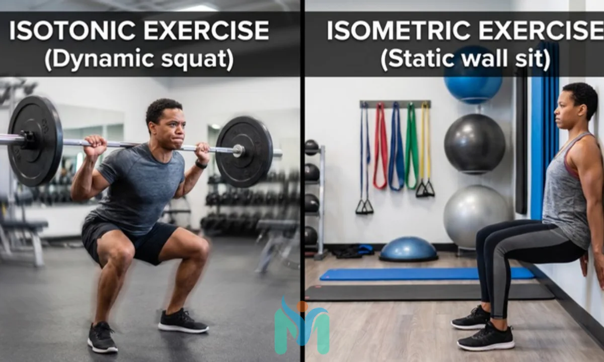
Isotonic and isometric exercise serve different purposes in physiotherapy, and the choice between them follows the stage of tissue healing and the clinical goal at that moment. Isometric exercise generates force without joint movement, making it appropriate earliest in recovery: directly after surgery, during acute inflammatory phases, or when mechanical loading of a joint is contraindicated. It maintains muscle activation and slows disuse atrophy without placing tensile or compressive stress on damaged structures.
Dynamic resistance work requires full joint motion and produces loading across the movement range. It is introduced as tissue integrity improves and the clinical priority shifts from protection to progressive functional loading. The movement demands produce a greater mechanical stimulus, which drives stronger adaptation but also requires tissue capable of tolerating that load without breakdown.
In practice, most rehabilitation programmes sequence these two types rather than choosing between them. Isometric work gets the neuromuscular system firing again and provides early pain management through motor unit recruitment. Isotonic loading follows, beginning at low loads and high repetitions, advancing progressively toward functional strength. Students who treat this as a binary choice often miss the transitional staging that defines competent rehabilitation planning.
Open and Closed Kinetic Chain Exercise: The Clinical Framework Behind Exercise Selection
Within isotonic exercise, a second classification system shapes prescription decisions: whether the movement is performed in an open kinetic chain (OKC) or a closed kinetic chain (CKC) configuration. This distinction affects muscle recruitment patterns, joint loading profiles, and how well the exercise translates to functional daily demands.

In OKC exercises, the distal segment (foot or hand) moves freely through space. A seated knee extension, a supine straight leg raise, or a shoulder external rotation with a resistance band are OKC movements. These load specific muscles with relative isolation, making them useful when a targeted structure needs loading without demanding full limb co-ordination from a system that is not yet ready for it.
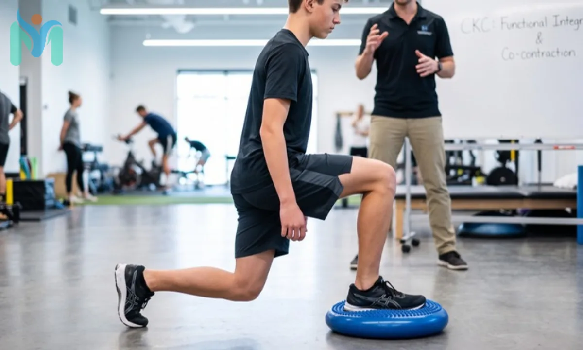
In CKC exercises, the distal segment is fixed against a surface while the body moves around the fixed point. Squats, step-ups, push-ups, and wall slides are CKC movements. A 1998 biomechanical study by Escamilla and colleagues in Medicine and Science in Sports and Exercise demonstrated that CKC knee exercises produce significantly greater co-contraction from the hamstrings and gastrocnemius compared to OKC equivalents, contributing to joint stability under load. This makes CKC exercises particularly appropriate for mid-to-late rehabilitation, where functional integration and joint confidence are the clinical priorities.
The clinical rule is clean: OKC exercises offer precision; CKC exercises offer function. Most rehabilitation programmes use both, sequenced by the structures being targeted and the stage of recovery.
Why Eccentric Exercise Changes the Rehabilitation of Tendons
Tendons adapt more slowly than muscle. They have limited blood supply and depend on mechanical loading to stimulate collagen synthesis and structural remodelling. A review by Kjaer in Physiological Reviews (2004) established that tendon collagen synthesis is directly stimulated by mechanical tensile load, and that load magnitude influences the quality of the tissue response. This is why the type of contraction matters as much as the load itself when rehabilitating tendinopathy.
Eccentric loading is the most thoroughly studied isotonic modality for tendon rehabilitation. The proposed mechanisms include stimulation of tenocyte activity (the cells responsible for tendon maintenance and repair), increased type I collagen synthesis, and reduction of pathological neovascularisation (the abnormal blood vessel ingrowth associated with tendon pain). A 2014 systematic review by Frizziero and colleagues in the British Medical Bulletin concluded that eccentric exercise is supported by consistent controlled evidence for Achilles and patellar tendinopathy, and should be considered a primary rehabilitation approach for these conditions.
The landmark clinical evidence came from a 1998 randomised controlled trial by Alfredson and colleagues in the American Journal of Sports Medicine. Patients with chronic Achilles tendinopathy who completed a 12-week heavy eccentric calf-loading protocol reported significant pain reduction and return to sport. The protocol has since been replicated across multiple trials. The following is how it is structured clinically, presented as an educational example of eccentric isotonic loading applied to tendinopathy:
Purpose: To apply high tensile load to the Achilles tendon during the lengthening phase of a calf contraction, stimulating tenocyte activity and type I collagen synthesis. The eccentric lengthening phase produces a tensile stimulus that concentric loading at the same resistance does not replicate.
Starting position: Stand on the edge of a step with the heel clear of the step edge, weight distributed through the forefoot of the affected limb. Rest one hand lightly on a wall or handrail for balance. Perform with the knee fully straight to target the gastrocnemius (the large, superficial calf muscle), and with the knee slightly bent to preferentially load the soleus (the deeper calf muscle with the most direct attachment into the Achilles tendon).
Movement: Use both limbs or the unaffected limb to rise to the top position (the concentric phase, which is not the therapeutic component). Transfer all body weight to the affected limb. Then slowly lower the heel below the level of the step over 3 to 4 seconds, allowing the calf musculature to lengthen fully under load. This controlled lowering is the therapeutic eccentric component. Return to the top using both limbs to begin the next repetition.
Dosage: 3 sets of 15 repetitions, twice daily, 7 days per week, for 12 weeks, as per the original Alfredson protocol. Mild to moderate tendon discomfort during exercise is expected within this protocol and does not indicate harm. The expected working discomfort is a deep, localised tendon ache that fades within 24 hours of a session. If discomfort does not resolve within that window, load may need to be reduced before progression resumes.
Safety note: Stop immediately if you experience a sharp, sudden pain clearly distinct from the expected tendon ache, or any sensation of tearing or sudden loss of tension in the calf. This protocol is designed for chronic Achilles tendinopathy and is not appropriate during acute reactive presentations or where tendon rupture is suspected.
A 2015 randomised controlled trial by Beyer and colleagues in the American Journal of Sports Medicine compared the Alfredson eccentric protocol to heavy slow resistance training in Achilles tendinopathy and found both equally effective at 12 weeks. A 2009 trial by Kongsgaard and colleagues in the Scandinavian Journal of Medicine and Science in Sports found heavy slow resistance training superior to eccentric loading alone in patellar tendinopathy at 6-month follow-up. The pattern across this evidence points to high mechanical load delivered through the isotonic range as the active ingredient, with eccentric loading being a particularly effective way to deliver it.
How Isotonic Exercise Is Dosed: Evidence-Based Load and Progression
Isotonic exercise dosage is structured around the specific adaptation being targeted. The foundational principle is progressive overload: the training stimulus must be systematically increased to continue driving adaptation, because the body adapts to a given load and stops responding to it. The principle was formalised for clinical rehabilitation by DeLorme in 1945, who developed the first structured progressive resistance exercise protocol. The American College of Sports Medicine’s 2009 position stand subsequently codified specific dosage parameters across training goals, providing the evidence base that modern physiotherapy prescription draws from.
| Training Goal | Load (% of 1RM) | Repetitions per Set | Sets | Rest Between Sets |
|---|---|---|---|---|
| Muscular endurance | Below 67% | 15 to 25 | 2 to 3 | 30 to 60 seconds |
| Hypertrophy (muscle size) | 67 to 85% | 8 to 12 | 3 to 5 | 60 to 90 seconds |
| Maximum strength | Above 85% | 2 to 6 | 3 to 6 | 2 to 5 minutes |
| Power development | 30 to 60% | 3 to 8 (explosive velocity) | 3 to 5 | 2 to 3 minutes |
The 1RM (one-repetition maximum: the heaviest load a person can lift for one complete repetition with correct technique) anchors load prescription. In early rehabilitation, direct 1RM testing is often clinically inappropriate, so load is estimated from submaximal repetition tests or repetitions performed to technical failure. Clinicians adjust load frequently, particularly in the first 4 to 8 weeks when neural adaptations drive rapid strength gains that quickly outpace the original prescription.
In practice, the most consistent error in isotonic exercise prescription is not loading too heavily: it is not loading enough, or not loading for long enough, to drive real physiological change. Under-loaded exercise protects healing tissue from excess stress but also fails to provide the stimulus needed for adaptation. The clinical skill is finding and progressively advancing the load that sits above the adaptation threshold without exceeding the tissue’s capacity to recover between sessions.
What Physiological Adaptations Does Isotonic Exercise Produce?
The adaptations from isotonic resistance exercise follow a predictable two-phase sequence. In the first 4 to 8 weeks, strength improvements are driven primarily by neural adaptations: the nervous system becomes more efficient at recruiting motor units (each a motor neuron and the muscle fibres it controls), synchronising their firing, and reducing inhibitory signals that previously limited force output. This is why people gain measurable strength early in a new programme before any visible change in muscle size has occurred.
After approximately 8 to 12 weeks of consistent progressive loading, morphological adaptations become the dominant driver of continued strength gains. Muscle fibres increase in cross-sectional area (hypertrophy), with Type II fast-twitch fibres showing the greatest size response to heavy resistance work. A 2010 review by Schoenfeld in the Journal of Strength and Conditioning Research identified 3 primary mechanisms driving hypertrophy: mechanical tension at moderate-to-high loads, metabolic stress from accumulated exercise metabolites, and muscle damage followed by repair-driven growth. Both concentric and eccentric isotonic contractions activate all 3 mechanisms, with relative contributions varying by load, volume, and contraction tempo.
At the connective tissue level, a 2014 review by Vogt and Hoppeler in the Journal of Applied Physiology confirmed that eccentric isotonic loading produces distinct molecular adaptations in both muscle and connective tissue, including changes in titin expression and collagen remodelling, that are not fully replicated by concentric-dominant training alone. The 2009 systematic review by Roig and colleagues further confirmed that eccentric-focused protocols produce greater Type II fibre hypertrophy than concentric-only approaches.
The specific adaptations produced by isotonic resistance exercise include:
- Increased muscle strength through improved motor unit recruitment and firing synchronisation in the first 8 weeks, with fibre hypertrophy taking over as the sustained driver beyond that point
- Greater cross-sectional area in both Type I slow-twitch and Type II fast-twitch muscle fibres, with Type II fibres showing a greater hypertrophic response to eccentric-dominant loading per the findings of Roig and colleagues (2009)
- Improved tendon stiffness and collagen density, which increases the efficiency of force transmission from muscle to bone and reduces injury risk under sustained high-load demand
- Enhanced neuromuscular co-ordination and inter-muscular timing, particularly relevant in CKC compound exercises where multiple joints must load in correct sequence
- Increased bone mineral density at sites of muscle attachment, a clinically meaningful benefit in older populations and those at elevated risk of osteoporosis
- Delayed onset muscle soreness in the 24 to 48 hours following a session, most pronounced after eccentric loading in untrained individuals, representing an acute adaptive inflammatory response rather than structural tissue damage in most presentations
Isotonic Exercise in Practice: Why Type, Kinetic Chain, and Load All Matter
Isotonic exercise is not a single prescription. It is a category that spans from gentle, low-load endurance work in early post-surgical rehabilitation to high-intensity maximum strength training for return to sport. What makes it central to physiotherapy is the underlying logic: a muscle dynamically loaded through a full range, at an appropriate intensity, adapts in ways that rest and passive treatment alone cannot achieve.
For students preparing for clinical placement, the most useful shift is from asking “which exercise?” to asking “which contraction type, in which kinetic chain configuration, at which load, for which stage of this person’s recovery?” A bicep curl and a push-up are both isotonic exercises. They deliver entirely different stimuli to different structures and are not interchangeable. The same movement performed at 60% of 1RM for 15 repetitions produces a different adaptation than at 85% for 6 repetitions in the same muscle.
There is one further clinical dimension that rarely surfaces clearly in introductory accounts of isotonic exercise: the importance of matching contraction type to the specific mechanical demands of the patient’s goal activity. A runner regularly descending hills places primarily eccentric demand on the quadriceps and calf complex. A rehabilitation programme that builds concentric quadriceps strength but does not specifically train eccentric capacity will not adequately prepare that runner for the actual loads of their sport. Prescribing isotonic exercise well means understanding not just what the general evidence supports, but what the specific individual in front of you needs to be able to do.
The evidence base for isotonic exercise in physiotherapy rehabilitation is among the strongest in the field. The American College of Sports Medicine has codified dosage parameters, the British Journal of Sports Medicine has confirmed eccentric loading superiority for hypertrophy and tendon rehabilitation, and multiple randomised controlled trials have demonstrated clinical outcomes across tendinopathy, post-surgical recovery, and musculoskeletal pain management. Converting that evidence into specific, well-reasoned, progressive prescriptions for individual patients is the task that defines clinical competence.
Frequently Asked Questions (FAQs)
Isotonic exercise is dynamic resistance exercise in which a muscle generates force while changing length against a constant external load. It encompasses concentric contractions, where the muscle shortens against resistance, and eccentric contractions, where it lengthens under load. According to the American College of Sports Medicine’s 2009 position stand on resistance training, progressive isotonic exercise produces measurable adaptations in muscular strength, hypertrophy, and endurance, making it the foundational training modality used throughout physiotherapy rehabilitation.
A concentric contraction occurs when a muscle shortens against resistance, overcoming the load to produce joint movement. An eccentric contraction occurs when the muscle lengthens under load, controlling resistance rather than overcoming it. Eccentric contractions generate greater peak force per cross-sectional area and a stronger hypertrophic stimulus, particularly in Type II fast-twitch fibres. In rehabilitation, eccentric loading is specifically targeted in tendinopathy protocols and in strengthening programmes where high tensile stimulus to the muscle-tendon unit is the clinical goal.
Eccentric exercise is recommended for tendinopathy because it applies high tensile load to the tendon during the lengthening phase of contraction, stimulating tenocyte activity and type I collagen synthesis. A 2014 systematic review by Frizziero and colleagues in the British Medical Bulletin confirmed consistent controlled evidence for eccentric loading in Achilles and patellar tendinopathy. The mechanical stimulus during eccentric lengthening promotes tendon collagen remodelling in ways concentric loading at equivalent resistance does not achieve.
Isotonic exercise involves dynamic muscle contraction through full joint range of movement against a constant resistance. Isometric exercise generates force with no change in muscle length and no joint movement. In physiotherapy, isometric exercise is used earliest in recovery when joint loading must be minimised, such as directly after surgery or during acute tissue irritability. Isotonic loading follows as healing permits, producing a stronger adaptive stimulus through dynamic loading of the muscle-tendon unit across its full functional range.
Open kinetic chain (OKC) exercises involve a free distal segment moving through space, as in a seated knee extension or a resistance band shoulder rotation. Closed kinetic chain (CKC) exercises fix the distal segment against a surface while the body moves around it, as in a squat or push-up. CKC exercises produce greater joint co-contraction and are more functionally relevant to daily activities and sport. OKC exercises allow more isolated muscle loading. Physiotherapy programmes typically use both, selected by clinical stage and the structures being targeted.
Dosage depends on the training goal. According to the American College of Sports Medicine’s 2009 position stand, muscular endurance requires lighter loads and 15 to 25 repetitions per set; hypertrophy requires moderate loads and 8 to 12 repetitions; maximum strength requires heavy loads and 2 to 6 repetitions. In rehabilitation, endurance parameters are introduced first to build tissue tolerance, with load progressively increased as strength improves. All dosage is calculated relative to the individual’s one-repetition maximum.
Consult your doctor or a qualified physiotherapist before starting any new exercise programme, especially if you have an existing injury or medical condition.
References
- American College of Sports Medicine. “American College of Sports Medicine position stand. Progression models in resistance training for healthy adults.” Medicine and Science in Sports and Exercise. 41(3):687–708 (2009). DOI: 10.1249/MSS.0b013e3181915670. PMID: 19204579. URL: https://pubmed.ncbi.nlm.nih.gov/19204579/. Evidence Level: 1.
- Roig M, O’Brien K, Kirk G, et al. “The effects of eccentric versus concentric resistance training on muscle strength and mass in healthy adults: a systematic review with meta-analysis.” British Journal of Sports Medicine. 43(8):556–568 (2009). DOI: 10.1136/bjsm.2008.051417. PMID: 18981046. URL: https://pubmed.ncbi.nlm.nih.gov/18981046/. Evidence Level: 2.
- Alfredson H, Pietilä T, Jonsson P, Lorentzon R. “Heavy-load eccentric calf muscle training for the treatment of chronic Achilles tendinosis.” American Journal of Sports Medicine. 26(3):360–366 (1998). PMID: 9617396. URL: https://pubmed.ncbi.nlm.nih.gov/9617396/. Evidence Level: 4.
- Frizziero A, Trainito S, Oliva F, et al. “The role of eccentric exercise in sport injuries rehabilitation.” British Medical Bulletin. 110(1):47–75 (2014). DOI: 10.1093/bmb/ldu006. PMID: 24736013. URL: https://pubmed.ncbi.nlm.nih.gov/24736013/. Evidence Level: 2.
- Kjaer M. “Role of extracellular matrix in adaptation of tendon and skeletal muscle to mechanical loading.” Physiological Reviews. 84(2):649–698 (2004). DOI: 10.1152/physrev.00031.2003. PMID: 15044685. URL: https://pubmed.ncbi.nlm.nih.gov/15044685/. Evidence Level: 5.
- Beyer R, Kongsgaard M, Hougs Kjær B, Øhlenschlæger T, Kjær M, Magnusson SP. “Heavy Slow Resistance Versus Eccentric Training as Treatment for Achilles Tendinopathy: A Randomized Controlled Trial.” American Journal of Sports Medicine. 43(7):1704–1711 (2015). DOI: 10.1177/0363546515584760. PMID: 25969018. URL: https://pubmed.ncbi.nlm.nih.gov/25969018/. Evidence Level: 4.
- Kongsgaard M, Kovanen V, Aagaard P, et al. “Corticosteroid injections, eccentric decline squat training and heavy slow resistance training in patellar tendinopathy.” Scandinavian Journal of Medicine and Science in Sports. 19(6):790–802 (2009). DOI: 10.1111/j.1600-0838.2009.00949.x. PMID: 19793213. URL: https://pubmed.ncbi.nlm.nih.gov/19793213/. Evidence Level: 4.
- Escamilla RF, Fleisig GS, Zheng N, Barrentine SW, Wilk KE, Andrews JR. “Biomechanics of the knee during closed kinetic chain and open kinetic chain exercises.” Medicine and Science in Sports and Exercise. 30(4):556–569 (1998). PMID: 9565938. URL: https://pubmed.ncbi.nlm.nih.gov/9565938/. Evidence Level: 4.
- Herzog W. “The role of titin in eccentric muscle contraction.” Journal of Experimental Biology. 217(16):2825–2833 (2014). DOI: 10.1242/jeb.099127. PMID: 25078283. URL: https://pubmed.ncbi.nlm.nih.gov/25078283/. Evidence Level: 5.
- Schoenfeld BJ. “The mechanisms of muscle hypertrophy and their application to resistance training.” Journal of Strength and Conditioning Research. 24(10):2857–2872 (2010). DOI: 10.1519/JSC.0b013e3181e840f3. PMID: 20847704. URL: https://pubmed.ncbi.nlm.nih.gov/20847704/. Evidence Level: 5.
- Vogt M, Hoppeler HH. “Eccentric exercise: mechanisms and effects when used as training regime or training adjunct.” Journal of Applied Physiology. 116(11):1446–1454 (2014). DOI: 10.1152/japplphysiol.00146.2013. PMID: 24505103. URL: https://pubmed.ncbi.nlm.nih.gov/24505103/. Evidence Level: 5.
- DeLorme TL. “Restoration of muscle power by heavy resistance exercises.” Journal of Bone and Joint Surgery (American Volume). 27:645–667 (1945). DOI: Not available (historical publication, pre-digital indexing). PMID: Not available (pre-PubMed era). Accessible via medical library database. Evidence Level: 6.