

That grinding sensation when you climb the stairs. The stiffness that locks your knee every time you stand after sitting for an hour. A dull ache that has become more familiar at night than it used to be. If any of this sounds like your knee, you are in company with hundreds of millions of people worldwide, and the research on knee osteoarthritis physiotherapy consistently points in a single direction: staying active, with the right guidance, is what works.
What Is Knee Osteoarthritis?
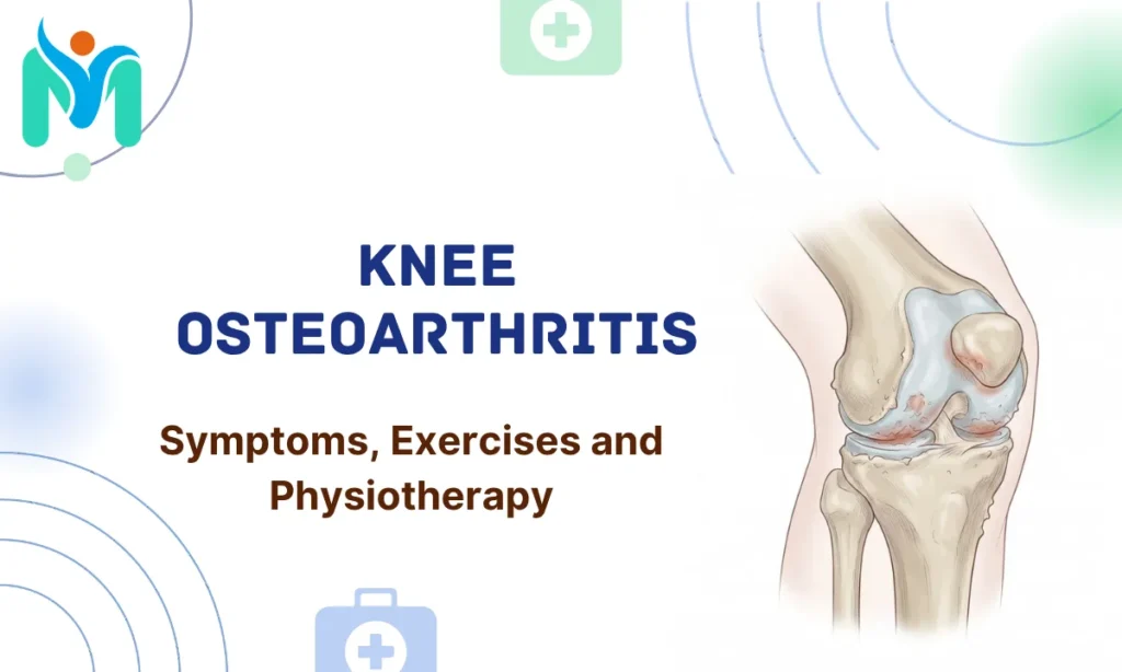
Knee osteoarthritis (OA) is a condition in which the articular cartilage (the smooth, slippery tissue covering the ends of the bones inside the knee joint) gradually breaks down. As this cartilage thins, the bones beneath begin to change: forming small bony outgrowths called osteophytes, thickening through a process known as subchondral bone sclerosis, and triggering inflammation in the surrounding joint lining called synovitis. The result is a joint that moves less freely, aches more reliably, and responds badly to the demands placed on it every day.
According to the World Health Organization, osteoarthritis affects approximately 528 million people globally, making it the most prevalent musculoskeletal condition in the world. The knee is the most frequently affected joint. In the UK, NHS guidance on osteoarthritis identifies it as one of the leading causes of pain and disability in adults over 45, and women are affected more often than men, particularly after the age of 50.
A number of factors increase the likelihood of developing knee OA. Age reduces the resilience of cartilage over time. Excess body weight increases the mechanical forces transmitted through the joint with every step. Previous knee injuries, occupations involving repeated kneeling or heavy lifting, and a family history of the condition all contribute. The condition is also known as degenerative joint disease, gonarthrosis, or wear-and-tear arthritis. These names all describe the same underlying process.
Knee osteoarthritis is a condition that responds directly to how you manage it. The research and clinical experience both tell a considerably more hopeful story than most people expect when they first receive the diagnosis.
Knee osteoarthritis is a degenerative joint condition in which the articular cartilage covering the surfaces of the femur (thigh bone), tibia (shin bone), and patella (kneecap) progressively breaks down. This produces pain, joint stiffness, swelling, reduced range of motion, and functional limitation. The World Health Organization estimates it affects hundreds of millions of people globally. It is the most common form of arthritis and a leading cause of disability in adults over 45 worldwide.
What Are the Stages of Knee Osteoarthritis?
Clinicians classify knee OA on X-ray using the Kellgren-Lawrence grading system, which describes the extent of joint changes from Grade 1 through to Grade 4. The grade on imaging gives one piece of the picture, and your physiotherapist will always combine it with a full clinical assessment to understand your actual capacity. Some people with Grade 3 changes function very well with physiotherapy; others with Grade 2 are significantly more limited in daily life. The table below gives a practical overview of what each grade typically means.
| Grade | What Imaging Shows | Typical Symptoms | Exercise Approach |
|---|---|---|---|
| Grade 1 – Doubtful | Minor osteophyte formation; joint space essentially normal | Occasional ache after prolonged activity; often no symptoms at rest | Full range of exercise; build long-term muscle strength |
| Grade 2 – Mild | Small osteophytes; slight joint space narrowing | Pain during or after activity; morning stiffness settling within 30 minutes | Progressive strengthening and aerobic exercise; increase load as tolerated |
| Grade 3 – Moderate | Multiple osteophytes; moderate joint space narrowing; early bone surface changes | Persistent pain; swelling; reduced range of motion; activity limitation | Modified loading; emphasise range of motion, hip strengthening, and aquatic exercise |
| Grade 4 – Severe | Large osteophytes; severe space narrowing; possible varus or valgus deformity | Constant pain; daily function substantially affected; possible visible deformity | Low-load and aquatic exercise; specialist physiotherapy assessment strongly recommended |
The grade confirms structural changes. What you can do, how far you progress, and how much function you recover depends on far more than a number on a scan.
Recognising the Symptoms of Knee Osteoarthritis
Knee OA symptoms can develop so gradually that many people adapt their lives around them before fully registering the extent of the change. Pain and stiffness are the most familiar features, but the full picture includes several presentations that catch people off guard. Crepitus (the grinding, clicking, or crunching sensation felt inside the knee during movement) is one of the most common. It is caused by irregular cartilage surfaces moving against each other, and while it can feel alarming, it does not reliably predict pain severity.
The symptoms that most commonly bring people into a physiotherapy clinic include:
- Deep, aching pain inside or around the knee, typically worse after sustained activity and better after rest in earlier stages
- Morning stiffness that eases within 30 minutes of moving (stiffness persisting longer than 30 minutes in the morning is more characteristic of inflammatory arthritis and warrants separate assessment)
- Swelling or a sensation of fullness in the joint, particularly after prolonged or unaccustomed activity
- Reduced range of motion, making it difficult to fully straighten or fully bend the knee
- Crepitus during movement, felt or heard as a grinding, clicking, or crunching inside the joint
- Pain and difficulty with weight-bearing activities such as stairs, rising from low chairs, or walking on uneven ground
- Occasional giving way or a feeling of instability, particularly when the quadriceps (the group of 4 muscles at the front of the thigh) has weakened considerably
What I see most often in clinic is that people have been quietly managing these symptoms for months, sometimes years, before seeking assessment. They have changed how they walk, given up activities they love, and assumed this is simply the price of getting older. The research consistently shows otherwise: active management produces meaningful improvements in pain, function, and quality of life for the vast majority of people with knee OA.
Pain at rest or at night is worth discussing with your physiotherapist or GP. It can occur in more advanced OA when the synovial membrane (the joint lining) is actively inflamed, and it may indicate a need for further investigation. A good clinical assessment also helps identify conditions that can mimic knee OA symptoms, including pes anserine bursitis (inflammation of a bursa on the inner side of the knee), meniscal degeneration, or pain referred from the hip or lumbar spine.
Why Exercise Is One of the Most Effective Treatments for Knee Osteoarthritis
The first instinct for many people with a painful knee is to rest it. That instinct makes sense, and for a short acute flare it has a place. Over weeks and months, though, avoiding movement makes knee OA considerably worse: muscles weaken, the joint stiffens, and pain becomes harder to manage rather than easier.
A 2015 Cochrane systematic review by Fransen and colleagues found that land-based exercise reduces pain and improves physical function in knee osteoarthritis with effect sizes comparable to non-steroidal anti-inflammatory drugs (NSAIDs). That places structured exercise alongside the most commonly used pharmacological treatment for OA pain and without the associated side effects. NICE guideline NG226 (2022) recommends therapeutic exercise as a core treatment for all adults with osteoarthritis, regardless of age, disease severity, or body weight. The 2019 ACR guidelines by Kolasinski and colleagues make an equally strong recommendation.
Articular cartilage has no direct blood supply: it depends entirely on the mechanical cycle of compression and release during movement to draw nutrients from the surrounding synovial fluid, making regular appropriate exercise one of the primary mechanisms through which cartilage receives the nutrition it needs to function.
The type of exercise matters too. A 2013 network meta-analysis published in the BMJ by Uthman and colleagues found that strengthening exercise produces the greatest reduction in pain, while aerobic and aquatic exercise each contribute to functional improvements. A 2014 systematic review and meta-regression by Juhl and colleagues confirmed a dose-response relationship: people who exercise more consistently gain greater pain relief and better function. A long-term systematic review by Pisters and colleagues (2007) confirmed that these benefits persist at 6 months and beyond, but only for people who maintain their exercise programme. The gains are sustainable with consistent effort.
Exercise helps knee osteoarthritis by strengthening the muscles that reduce joint load, improving cartilage nutrition through synovial fluid circulation, and activating neurological pain-modulating pathways that reduce chronic pain sensitivity. A 2015 Cochrane systematic review found that land-based exercise reduces knee OA pain with effects comparable to NSAIDs. NICE guideline NG226 (2022) and the 2019 American College of Rheumatology guidelines both strongly recommend exercise as a first-line core treatment for all adults with knee osteoarthritis, regardless of disease severity or grade.
5 Best Exercises for Knee Osteoarthritis
The 5 exercises below are ordered from least to most physiologically demanding. Begin at the start of the sequence, particularly if you are newly beginning rehabilitation or managing Grade 3 to 4 OA. A practical guide to pain during exercise: mild discomfort of 2 to 3 out of 10 on a pain scale is acceptable and does not signal harm. Pain above 4 out of 10, any sharp or sudden pain, or pain that persists longer than 30 minutes after finishing the session means stopping that exercise and discussing it with your physiotherapist before continuing.
These exercises are educational examples. A physiotherapist will select, adapt, and progress them based on your individual assessment findings and the grade of your OA.
1. Seated Heel Slide
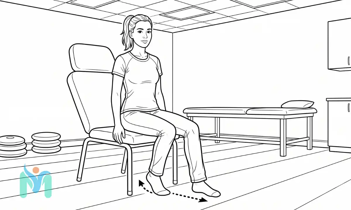
Purpose: Maintains and gently restores knee range of motion without placing compressive load through the joint surface. This exercise is particularly useful during flare-ups or for Grade 3 to 4 OA when weight-bearing exercise is poorly tolerated.
Start position: Sit in a firm, stable chair. Place a folded towel under your heel or wear socks to reduce friction on the floor beneath you.
Movement: Slowly slide your heel backward toward the chair legs, bending the knee as far as a comfortable, gentle stretch allows. Hold for 3 to 5 seconds. Slide the heel forward again until the knee is as straight as it will comfortably go. That is 1 repetition.
Dosage: 2 sets of 10 repetitions. Move deliberately throughout the full available range on every repetition.
Safety note: A gentle pulling sensation inside the knee is normal. Sharp or pinching pain at any point means reduce the range of motion and discuss it with your physiotherapist before proceeding.
2. Straight Leg Raise
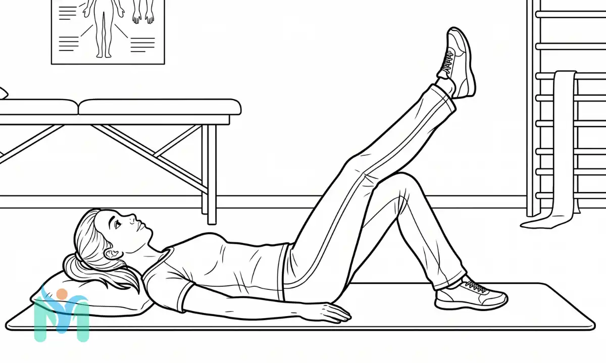
Purpose: Activates and strengthens the quadriceps without placing any compressive force through the knee joint. The quadriceps is the primary muscular shock absorber for the knee: when it weakens, mechanical stress transfers directly onto the articular cartilage and surrounding joint structures with every step taken.
Start position: Lie on your back on a firm, flat surface such as an exercise mat. Bend your unaffected knee to roughly 90 degrees with the foot flat on the mat. Keep the affected leg completely straight, with the foot gently pointed upward.
Movement: Tighten the thigh muscles of the straight leg until you feel the back of the knee press gently downward toward the mat. Lift the entire leg to the height of the opposite knee, approximately 30 centimetres off the surface. Hold for 2 seconds. Lower slowly and with full control. That is 1 repetition.
Dosage: 3 sets of 12 to 15 repetitions. Rest 60 seconds between sets.
Safety note: Keep the raised knee completely straight throughout the movement. If your lower back arches away from the mat, reduce the lift height slightly until your core control improves.
3. Clamshell
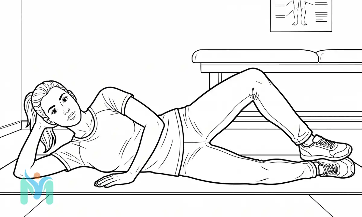
Purpose: Strengthens the hip abductors, particularly gluteus medius, which stabilise the pelvis and directly reduce the compressive forces acting through the medial (inner) compartment of the knee during walking. Hip abductor weakness is common in knee OA and amplifies the mechanical loading pattern on the damaged joint surface.
Start position: Lie on your side with your hips flexed to roughly 45 degrees and your knees stacked directly on top of each other. Keep your feet together throughout. A pillow beneath your head helps maintain a neutral spine position.
Movement: Keeping your feet touching, rotate your top knee upward toward the ceiling as far as is comfortable, without your top hip rolling backward. Lower with full control. Perform all repetitions on one side before switching.
Dosage: 3 sets of 12 to 15 repetitions on each side.
Safety note: The pelvis must remain completely still throughout. If your hips roll backward as you lift, reduce the range. All the movement should come from the hip rotating, with nothing from the pelvis itself.
4. Mini Wall Squat
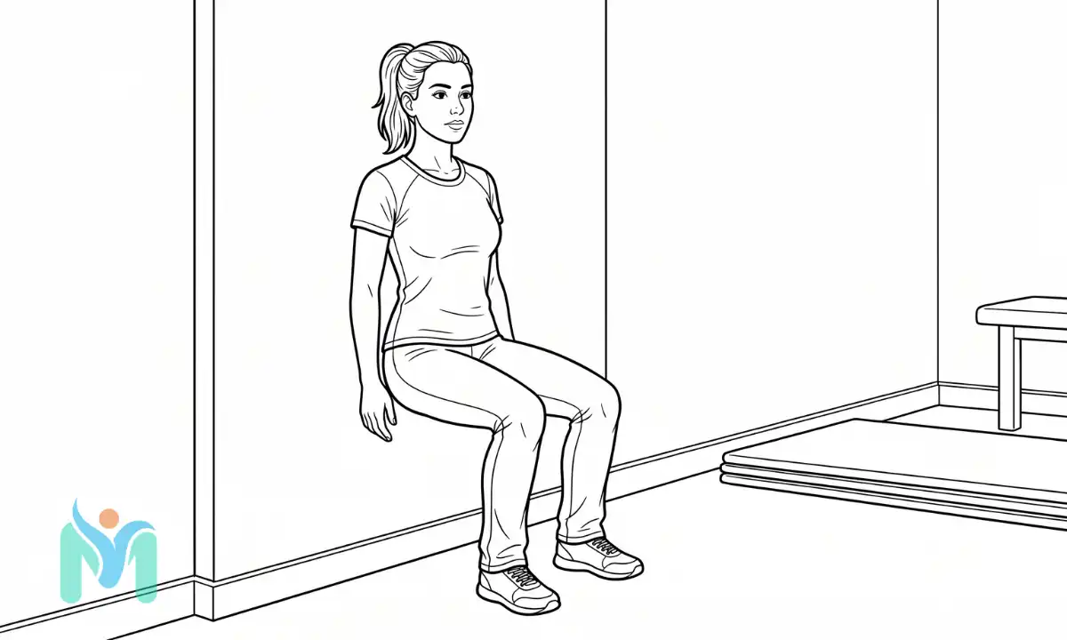
Purpose: Builds quadriceps strength through a closed-chain movement (where the foot stays in contact with the floor), which closely mirrors the muscle activation needed to rise from a chair, descend stairs, or walk on an incline. A 2014 systematic review by Juhl and colleagues confirmed that strengthening exercise of this type produces dose-dependent reductions in knee OA pain and disability.
Start position: Stand with your back flat against a smooth wall. Place your feet roughly 30 centimetres out from the wall, shoulder-width apart.
Movement: Slowly slide your back down the wall until your knees reach about 30 to 40 degrees of bend. Your thighs should feel the effort, but the knees should remain behind your toes. Hold the position for 5 to 10 seconds. Slide back up to full standing. That is 1 repetition.
Dosage: 3 sets of 8 to 10 repetitions. As strength improves, progress by increasing the hold time toward 20 seconds, or deepening the squat gradually toward 60 degrees over several weeks.
Safety note: Keep your knee aligned over your second toe at all times. Stop if you feel sharp pain at the kneecap (patellofemoral pain). This exercise places more demand than the earlier two, so introduce it only once those feel comfortable.
5. Step-Up
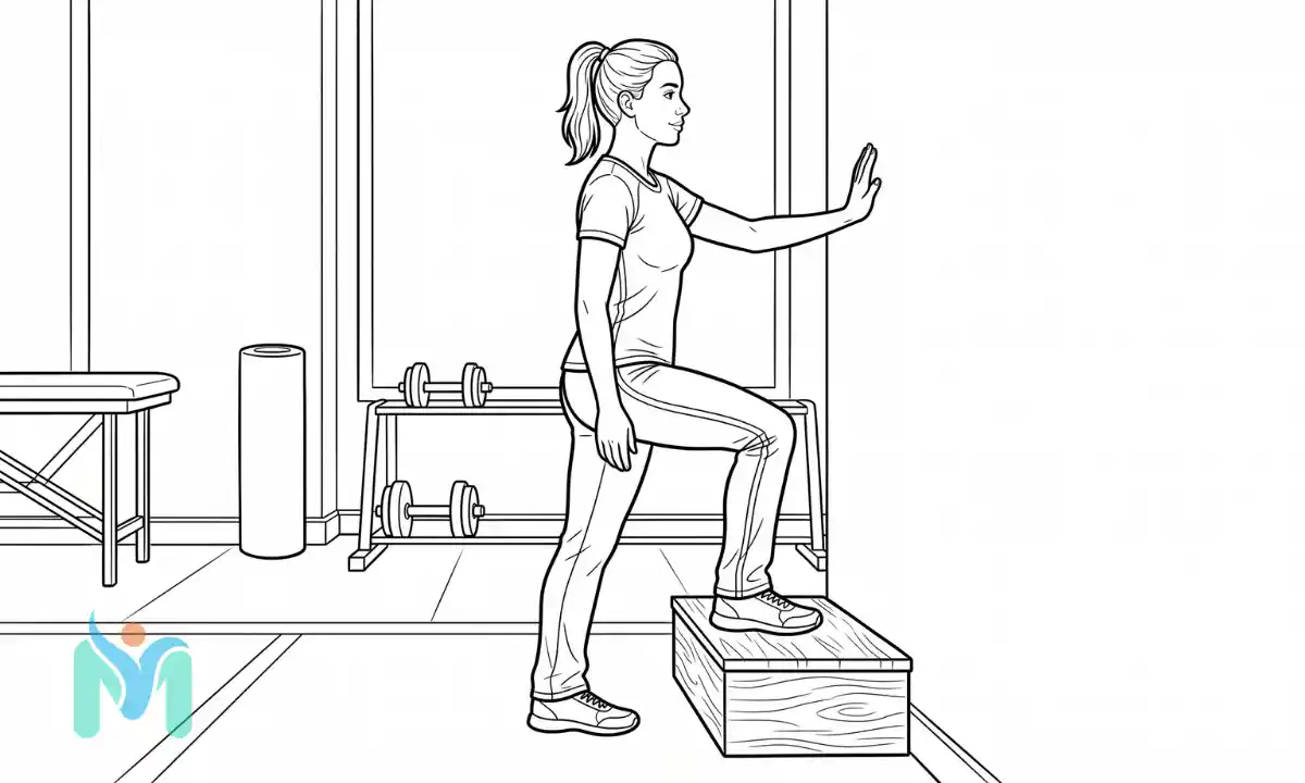
Purpose: Trains the quadriceps and gluteal muscles through a full weight-bearing, functional movement pattern that prepares the knee for real-world demands such as stair climbing, stepping off a kerb, or getting in and out of a vehicle. This is the most demanding exercise in this sequence and should only be introduced once the previous 4 feel comfortable and pain-free.
Start position: Stand in front of a low, stable step or platform 10 to 15 centimetres in height. Use a wall or handrail for balance as needed.
Movement: Step up with the affected leg leading, pressing through that heel to lift your body weight. Bring the other foot to join it on the step. Step back down leading with the unaffected leg first. Concentrate on the descent: lowering slowly and with control builds strength through the eccentric (lengthening) phase of the muscle contraction, which is where much of the functional value lies.
Dosage: 3 sets of 8 to 10 repetitions per leg.
Safety note: Keep the stepping knee tracking over the second toe at all times and control the speed of the descent. If knee pain increases during the exercise or lingers for more than 30 minutes afterward, return to the earlier exercises and rebuild before reintroducing step-ups.
What Does a Physiotherapist Do for Knee Osteoarthritis?
A physiotherapist treating knee OA does considerably more than provide a list of exercises. The clinical assessment examines how the knee moves, which muscles are underperforming, what your walking pattern reveals about the forces going through the joint, and how your hip and foot mechanics may be contributing. Knee OA rarely exists in isolation from what is happening above and below it.
A physiotherapist treating knee osteoarthritis carries out a detailed assessment of joint mobility, muscle strength, gait mechanics, and functional capacity. Treatment combines individually prescribed therapeutic exercise, manual therapy (skilled mobilisation of the joint and surrounding soft tissue), patient education, and activity modification guidance. A randomised controlled trial by Deyle and colleagues (2000) found that combining manual physical therapy with exercise produced considerably greater improvements in knee OA outcomes than exercise alone at both 8 weeks and 1 year. NICE guideline NG226 (2022) recommends physiotherapy as a central component of core management for all adults with osteoarthritis. Outcome measures including the KOOS (Knee injury and Osteoarthritis Outcome Score) and WOMAC (Western Ontario and McMaster Universities Arthritis Index) are used to track pain, stiffness, and function objectively throughout the programme.
Manual therapy for knee OA involves skilled, graded movements applied to the joint to improve range of motion and reduce pain. Your physiotherapist may also address the soft tissues around the knee: the quadriceps, the iliotibial band (the thick connective tissue running along the outer thigh), the calf complex, and the hip musculature. These techniques work on the muscular and movement contributions to pain rather than on the cartilage itself, and they can reduce the muscle guarding, restricted movement patterns, and compensatory gait changes that often amplify symptoms considerably.
Physiotherapy also addresses activity pacing and load management. Understanding how to structure your day to protect the knee during reactive periods, when supportive equipment such as bracing or orthotic insoles is appropriate, and how to progress your exercise programme at the right rate are all part of what good physiotherapy for knee OA delivers.
What I see most often in clinic is that people arrive expecting to be told to rest. Effective knee osteoarthritis physiotherapy is almost entirely the opposite: it is active, progressive, and requires real commitment. The 2019 OARSI guidelines by Bannuru and colleagues, the 2019 ACR guidelines, and NICE NG226 are all aligned on this point: exercise-based physiotherapy is a first-line recommendation for every grade of knee OA.
For Grade 3 to 4 OA, or when symptoms have not responded to well-supervised conservative management over 3 to 6 months, your physiotherapist may discuss onward referral. Options can include corticosteroid injections to reduce joint inflammation during a pronounced flare, hyaluronic acid injections, or orthopaedic review for consideration of unicompartmental or total knee replacement. These decisions are made collaboratively with your GP and specialist, and your physiotherapist plays an important role in that conversation.
Which Exercises and Activities to Approach With Care
Exercise is strongly and consistently recommended for knee OA. That recommendation covers the broad category of appropriate physical activity, and it does not mean every type of exercise is equally well-suited at every stage or for every individual. Some activities place considerably higher compressive or shear forces through a damaged joint surface, particularly when the muscular support around the knee is still limited at the start of rehabilitation. Most people eventually return to activities they have previously reduced, but doing so works best when the muscular foundation has been established first.
- Deep squats and lunges (beyond 90 degrees of knee bend) load the patellofemoral joint and tibiofemoral cartilage substantially, particularly on weakened or asymmetrical musculature
- Running on hard surfaces in the early stages of rehabilitation, before adequate quadriceps and hip strength has been built, can aggravate symptoms and slow recovery
- High-impact activities involving jumping, rapid direction changes, or cutting movements, which generate forces the joint cannot yet absorb well
- Prolonged kneeling directly on the knee, which can irritate the prepatellar or infrapatellar bursae (the small fluid-filled structures that cushion the front of the joint)
- Extended periods of continuous standing on hard flooring without rest breaks, particularly during the early reactive phase
- Continuing any exercise through pain above 3 to 4 out of 10 on a pain scale, or any activity that causes pain persisting more than 30 minutes after stopping
Aquatic exercise is an excellent alternative when land-based activity is poorly tolerated. Water reduces the effective body weight loading through the knee by up to 75 per cent depending on water depth, while still allowing meaningful muscle activation and range of motion work. A 2016 Cochrane systematic review by Bartels and colleagues found that aquatic exercise produces meaningful reductions in knee OA pain and improves quality of life with a strong safety profile across the available evidence.
Weight management is worth noting separately. The ADAPT trial, a large randomised controlled trial by Messier and colleagues (2004), found that combining dietary weight loss with exercise produced considerably greater reductions in knee OA pain and disability than either approach alone in overweight older adults. Reducing body weight reduces the mechanical load transmitted through the joint surface on every step, and it is one of the most evidence-grounded self-management strategies available to those above a healthy weight range.
Frequently Asked Questions (FAQs)
Physiotherapy cannot reverse the structural changes of knee osteoarthritis, including cartilage loss or osteophyte formation. What a well-designed programme consistently achieves is a meaningful reduction in pain, improvements in muscle strength, better joint mobility, and improved daily function. Multiple systematic reviews and clinical guidelines confirm this. Many people with confirmed knee OA achieve excellent long-term outcomes through structured physiotherapy and sustained exercise without requiring surgery.
Most people notice some reduction in pain and improvement in function within 6 to 8 weeks of a consistent exercise programme. Meaningful functional gains typically emerge over 3 to 6 months of regular effort. A 2007 systematic review by Pisters and colleagues found that exercise benefits in knee OA are maintained at 6 months and beyond for people who keep exercising. Short treatment courses followed by stopping exercise tend to result in symptoms returning within months.
Walking is generally beneficial and actively recommended by NICE guideline NG226 (2022) and the 2019 ACR guidelines for adults with knee OA. It supports aerobic fitness, assists with weight management, maintains joint range of motion, and contributes to cartilage nutrition through regular movement. Begin with comfortable distances on flat, even surfaces and build gradually. If walking triggers pain above 4 out of 10 that does not settle within 30 minutes of stopping, reduce the distance and discuss modification with a physiotherapist.
A 2013 network meta-analysis published in the BMJ by Uthman and colleagues found that strengthening exercises produce the greatest pain reduction in knee osteoarthritis. Aquatic and aerobic exercise each provide functional benefits. The 2019 OARSI guidelines by Bannuru and colleagues strongly recommend both land-based and aquatic exercise. In practice, the strongest programmes combine quadriceps and hip strengthening with aerobic activity, progressed according to individual ability and OA grade.
During a pronounced flare, when the knee is swollen, noticeably warmer than usual, or considerably more painful, reducing exercise intensity temporarily is reasonable. Low-load movements such as seated heel slides, gentle swimming, or walking in water are usually still appropriate and help prevent muscle loss during the flare. Stopping all exercise for extended periods worsens muscle weakness and overall joint function. A physiotherapist can modify your programme during a flare rather than suspending it.
Most people with knee OA manage well with conservative treatment and do not need surgery. A surgical opinion is typically considered when pain is severe and persistent despite 3 to 6 months of consistent, well-supervised management, when daily function is substantially impaired, and when imaging confirms Grade 3 to 4 changes alongside clear clinical limitation. Arthritis UK provides accessible information on knee replacement for those exploring this option. Your GP and physiotherapist can guide any referral process.
Staying Active Is the Best Thing You Can Do for Your Knee
Knee osteoarthritis responds directly to how you manage it. The evidence across decades of trials, systematic reviews, and clinical guidelines is consistent: exercise is the treatment for knee OA, and knee osteoarthritis physiotherapy is the framework that helps you do it well. A 2007 systematic review by Pisters and colleagues confirmed that exercise benefits persist at 6 months and beyond for people who keep exercising after a physiotherapy programme. Those who stop lose the gains, usually within months.
What this means practically is that the goal of physiotherapy for knee OA is not to deliver a fixed course of treatment and return you to the same habits. It is to give you the knowledge, the muscle strength, and the movement confidence to keep the joint working at its best for as long as possible. The people who do best with knee OA long term are consistent, they understand why the work matters, and they adapt their programme intelligently as their circumstances change.
Pain is a signal worth interpreting carefully. Understanding what it communicates is more clinically useful than defaulting to complete rest. Stiffness that builds in a resting knee is the joint’s way of asking to move. For most people, a knee OA diagnosis is the beginning of a more intentional approach to an active life, not the end of one. That is what the research supports, and it is what good physiotherapy makes possible.
Consult your doctor or a qualified physiotherapist before starting any new exercise programme, especially if you have an existing injury or medical condition.
References
- World Health Organization. “Osteoarthritis.” WHO Fact Sheet. https://www.who.int/news-room/fact-sheets/detail/osteoarthritis (2023).
- NHS. “Osteoarthritis.” NHS Conditions. https://www.nhs.uk/conditions/osteoarthritis/ (2023).
- Fransen M, McConnell S, Harmer AR, Van der Esch M, Simic M, Bennell KL. “Exercise for osteoarthritis of the knee.” Cochrane Database of Systematic Reviews. 2015;1:CD004376. PMID: 25569281. URL: https://pubmed.ncbi.nlm.nih.gov/25569281/.
- Kolasinski SL, Neogi T, Hochberg MC, et al. “2019 American College of Rheumatology/Arthritis Foundation Guideline for the Management of Osteoarthritis of the Hand, Hip, and Knee.” Arthritis Care and Research. 2020;72(2):149-162. PMID: 31908149. URL: https://pubmed.ncbi.nlm.nih.gov/31908149/.
- Uthman OA, van der Windt DA, Jordan JL, et al. “Exercise for lower limb osteoarthritis: systematic review incorporating trial sequential analysis and network meta-analysis.” BMJ. 2013;347:f5555. PMID: 24055922. DOI: 10.1136/bmj.f5555. URL: https://pubmed.ncbi.nlm.nih.gov/24055922/.
- Juhl C, Christensen R, Roos EM, Zhang W, Lund H. “Impact of exercise type and dose on pain and disability in knee osteoarthritis: a systematic review and meta-regression analysis of randomized controlled trials.” Arthritis and Rheumatology. 2014;66(3):622-636. PMID: 24574223. URL: https://pubmed.ncbi.nlm.nih.gov/24574223/.
- Pisters MF, Veenhof C, van Meeteren NL, et al. “Long-term effectiveness of exercise therapy in patients with osteoarthritis of the hip or knee: a systematic review.” Arthritis and Rheumatism. 2007;57(7):1245-1253. PMID: 17907210. URL: https://pubmed.ncbi.nlm.nih.gov/17907210/.
- Deyle GD, Henderson NE, Matekel RL, et al. “Effectiveness of manual physical therapy and exercise in osteoarthritis of the knee: a randomized, controlled trial.” Annals of Internal Medicine. 2000;132(3):173-181. PMID: 10651597. URL: https://pubmed.ncbi.nlm.nih.gov/10651597/.
- Bannuru RR, Osani MC, Vaysbrot EE, et al. “OARSI guidelines for the non-surgical management of knee, hip, and polyarticular osteoarthritis.” Osteoarthritis and Cartilage. 2019;27(11):1578-1589. PMID: 31278997. URL: https://pubmed.ncbi.nlm.nih.gov/31278997/.
- Bartels EM, Juhl CB, Christensen R, et al. “Aquatic exercise for the treatment of knee and hip osteoarthritis.” Cochrane Database of Systematic Reviews. 2016;3:CD005523. PMID: 27007113. URL: https://pubmed.ncbi.nlm.nih.gov/27007113/.
- Messier SP, Loeser RF, Miller GD, et al. “Exercise and dietary weight loss in overweight and obese older adults with knee osteoarthritis: the Arthritis, Diet, and Activity Promotion Trial.” Arthritis and Rheumatism. 2004;50(5):1501-1510. PMID: 15146420. URL: https://pubmed.ncbi.nlm.nih.gov/15146420/.
- Arthritis UK. “Osteoarthritis.” Arthritis UK. https://www.arthritis-uk.org/information-and-support/understanding-arthritis/conditions/osteoarthritis/ (2023).