
Your shoulder stopped cooperating weeks ago, and you’ve been quietly working around it: reaching with your other arm, avoiding the overhead shelf, figuring out new ways to fasten a seatbelt. The pain wakes you at 3 a.m. when you roll onto it. Then someone says “frozen shoulder” and tells you to wait.
Waiting without understanding what to do is the worst version of this situation. Frozen shoulder exercises are the primary tool for recovery, but the exercises that help in one stage of this condition can genuinely worsen symptoms in another. This guide explains which exercises suit each stage and why the order matters as much as the movements themselves.
What actually happens when a shoulder freezes?
Frozen shoulder, medically known as adhesive capsulitis (a thickening and contraction of the connective tissue surrounding the shoulder joint), affects 2 to 5% of the general population. According to a 2005 review published in the BMJ by Dias, Cutts, and Massoud, the condition progresses through 3 predictable clinical stages and typically resolves over 1 to 3 years, though some people retain long-term restriction without appropriate management.
The glenohumeral joint (the ball-and-socket joint where the rounded head of your upper arm bone meets the shallow socket of your shoulder blade) sits inside a sleeve of connective tissue called the joint capsule. In a healthy shoulder, this capsule is loose enough to accommodate your arm’s wide arc of movement. In frozen shoulder, the capsule undergoes two overlapping processes: first, the synovial membrane (the capsule’s smooth inner lining) becomes inflamed and thickened; then fibroblasts (cells responsible for laying down connective tissue) deposit excessive collagen, hardening the capsule into a contracted state that cannot release on its own.
A 2010 review by Neviaser and Hannafin in the American Journal of Sports Medicine put precise numbers to this process: the internal volume of the glenohumeral joint can shrink from its normal 20 to 30 millilitres to as little as 5 millilitres as adhesive capsulitis progresses. Two structures drive this contraction: the coracohumeral ligament (a band of tissue running between the coracoid process at the front of your shoulder blade and the top of your humerus) and the axillary recess (the lower fold of the joint capsule), both of which thicken and shorten over time.
This is why external rotation (turning your palm to face forward while your arm hangs at your side) and overhead reach disappear first. The coracohumeral ligament and anterior joint capsule are responsible for those movements, and they contract earliest in the condition’s progression.
Frozen shoulder, or adhesive capsulitis, is a condition in which the glenohumeral joint capsule becomes inflamed and gradually replaced by dense fibrous scar tissue, severely contracting the joint and restricting movement in all directions. According to NHS guidance and the 2005 BMJ review by Dias, Cutts, and Massoud, it affects 2 to 5% of the general population and typically resolves over 1 to 3 years. Physiotherapy exercise has been shown to improve functional recovery during this period.
The three stages of frozen shoulder: what each one means for your exercises
Each stage of frozen shoulder brings a different symptom pattern and responds best to a different type of exercise. Understanding which stage you’re in changes what you should be doing, not just how much effort you apply.
The classic 3-stage model was first described by Reeves in a 1975 longitudinal study of frozen shoulder outcomes, and it remains the basis for the exercise strategies endorsed in the 2012 evidence-based clinical practice guideline from the Chartered Society of Physiotherapy, led by Hanchard and colleagues.
| Stage | Name | Typical duration | Main feature | Exercise focus |
| 1 | Painful (Freezing) | 2 to 9 months | Severe pain, often worst at night; stiffness beginning | Gentle, pain-free movement within tolerance |
| 2 | Adhesive (Frozen) | 4 to 12 months | Severe stiffness in all directions; pain reducing | Sustained stretching to progressively recover range |
| 3 | Resolution (Thawing) | 5 to 24 months | Movement gradually returning; pain settling | Active strengthening and functional rehabilitation |
Frozen shoulder progresses through 3 clinical stages. Stage 1 (Painful or Freezing, lasting 2 to 9 months) features severe pain that is often worst at night, with stiffness just beginning to emerge. Stage 2 (Adhesive or Frozen, 4 to 12 months) brings severe stiffness in all directions with reducing pain. Stage 3 (Resolution or Thawing, 5 to 24 months) sees movement gradually returning. Stage boundaries overlap and individual timelines vary considerably, so staging is best confirmed by a physiotherapist through clinical assessment.
Many people find their symptoms don’t sit neatly inside one column. If you’ve had 3 months of significant pain and recently noticed that stiffness is overtaking pain as the main problem, you’re most likely moving from Stage 1 into Stage 2. That shift in symptom pattern changes which exercises will produce the most progress.
Who gets frozen shoulder, and why diabetes changes the picture
Frozen shoulder most commonly develops between ages 40 and 60, with a slight female predominance and a tendency to affect the non-dominant arm. Beyond these baseline patterns, the link with certain health conditions is the detail most general resources understate.
People with diabetes face a substantially higher risk of developing frozen shoulder. A 2016 meta-analysis by Zreik, Malik, and Charalambous in Muscles, Ligaments and Tendons Journal found that between 10 and 20% of people with diabetes develop adhesive capsulitis, compared with 2 to 5% of the general population, and diabetic patients tend to experience a longer and less predictable recovery. Thyroid disorders, cardiovascular disease, previous shoulder immobilisation, and conditions affecting arm use also appear consistently as contributing risk factors across the medical literature.
Other factors associated with increased risk include:
- Thyroid disorders, both overactive (hyperthyroidism) and underactive (hypothyroidism), appear repeatedly in the research as predisposing conditions
- Cardiovascular disease and previous cardiac events, including recovery from heart surgery where arm movement is restricted
- Previous shoulder injury, surgery, or any period during which the arm was immobilised in a sling or cast
- Dupuytren’s contracture (a hand condition in which fibrous tissue in the palm thickens and draws fingers inward)
- Stroke or other neurological conditions that reduce voluntary shoulder movement over time
- Parkinson’s disease and similar conditions involving extended periods of reduced limb use
If you have diabetes and a frozen shoulder, tell your physiotherapist at the first appointment. It affects the expected timeline and can influence how the stretching programme is progressed over the months ahead.
Should you exercise a frozen shoulder? The pain question answered
Exercise is the primary treatment for frozen shoulder, not a complementary option to consider alongside other things. The 2012 evidence-based clinical practice guideline from the Chartered Society of Physiotherapy, led by Hanchard and colleagues, identifies physiotherapy exercise and mobilisation as an effective management approach across all stages of the condition. But there is a meaningful difference between the type of pain you should work through and the type that tells you to stop.
A gentle ache during a frozen shoulder stretch that settles within 30 minutes of finishing exercise is working pain: expected, therapeutic, and a sign the contracted capsule is responding to progressive load. Warning pain is sharp, sudden, worsening during movement, or persists more than 1 hour after exercise. Warning pain means stop, rest, and reduce the exercise intensity at the next session.
In Stage 1, the inflamed joint capsule is reactive. Forcing range of motion during this phase can provoke synovitis further and extend the painful stage. The goal in Stage 1 is gentle movement within the pain-free range, maintaining what mobility you have while the inflammatory process runs its course. In Stage 2, where inflammation has largely converted to fibrous contracture, sustained stretch with mild discomfort is expected and necessary: the contracted capsule needs time under tension to progressively lengthen.
Most people in Stage 1 find they can tolerate more movement in the morning after a warm shower than in the evening after a day of protective holding. That pattern is worth using: exercise when tissue is most receptive, keep movements gentle, and stay within the pain-free range until the Stage 2 stiffness pattern begins to dominate.
Frozen shoulder exercises: your stage-specific programme
Evidence-based frozen shoulder exercises vary by stage. Stage 1 exercises focus on maintaining joint mobility without provoking the inflamed capsule. Stage 2 exercises apply progressive sustained stretch to the contracted joint capsule. Stage 3 exercises rebuild active shoulder elevation and rotational strength. A 2013 clinical practice guideline from the Orthopaedic Section of the American Physical Therapy Association, developed by Kelley and colleagues and published in the Journal of Orthopaedic and Sports Physical Therapy, identifies this staged progression as the primary intervention approach for adhesive capsulitis.
A 2014 Cochrane review by Page, Green, and colleagues found that exercise combined with manual therapy produces clinically meaningful improvements in shoulder pain and function. The exercises below form the self-management component of that combined approach.
Stage 1 exercises: Painful (Freezing) stage
During Stage 1, the joint capsule is actively inflamed. These exercises maintain what movement you have and keep the joint nourished without provoking the inflammatory process. Keep all movements within the pain-free range. If any exercise produces pain lasting more than 30 minutes after stopping, reduce the range and try again the following day.
1. Pendulum (Codman) exercise
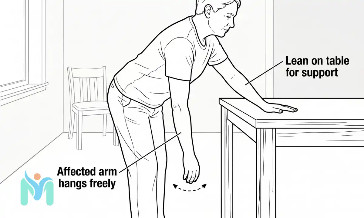
Purpose: The weight of your hanging arm creates a gentle tractioning force through the glenohumeral joint, reducing compressive load on the inflamed capsule. This encourages synovial fluid circulation without engaging the rotator cuff muscles, keeping the joint nourished without provoking inflammation.
Start position: Stand beside a table or sturdy chair. Lean forward at the hips and rest your unaffected arm flat on the surface for support. Let your affected arm hang completely relaxed, straight down toward the floor.
Movement: Using a gentle rock of your body, let the affected arm swing in small clockwise circles, driven by body momentum rather than any shoulder muscle effort. After 10 to 15 circles, reverse to anti-clockwise. The circles should be small, roughly 20 to 30 centimetres across. The shoulder does no active work.
Dosage: 10 to 15 circles in each direction, 2 to 3 times daily.
Safety note: If the shoulder tightens or you feel yourself actively lifting the arm, reduce circle size. This exercise should feel entirely passive throughout.
2. Supported table slide
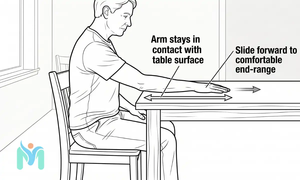
Purpose: The smooth table surface carries the full weight of your arm, allowing gentle shoulder elevation without rotator cuff engagement. This maintains the shoulder’s capacity to move into elevation while the joint is too inflamed to tolerate loaded movement, preventing the capsule from stiffening in a fixed position.
Start position: Sit at a smooth table or countertop. Place your affected arm flat on the surface, palm down, with your elbow straight.
Movement: Slide your arm slowly forward along the table, as if reaching toward the far edge. Let the table carry your arm’s weight throughout. Slide until you feel a gentle pull at the front of the shoulder, hold for 2 to 3 seconds, then slide back to start. The surface does the supporting work.
Dosage: 10 repetitions, 2 to 3 times daily. Gradually increase the sliding distance as comfort allows over subsequent sessions.
Safety note: Keep your arm in contact with the table surface at all times. Do not lift the arm off the table or push upward against gravity.
Stage 2 exercises: Adhesive (Frozen) stage
In Stage 2, the inflammatory process has largely converted to fibrous contracture. The capsule is stiff but less reactive than in Stage 1. Mild discomfort during sustained stretching is expected at this stage: the contracted tissue needs time under tension to progressively lengthen. Stop if you feel sharp or worsening pain at any point.
3. Cross-body horizontal stretch
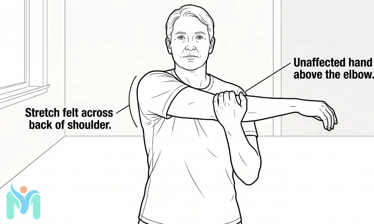
Purpose: The posterior joint capsule (the back wall of the shoulder capsule) contracts in Stage 2 and limits horizontal movement across the body. It also alters how the head of the humerus tracks within the glenoid socket, affecting the quality of every other shoulder movement. This stretch targets the posterior capsule specifically, restoring horizontal reach and improving the foundation for all other capsular stretching.
Start position: Stand or sit upright. Bring your affected arm horizontally across your chest at shoulder height.
Movement: Place your unaffected hand just above the elbow of the affected arm. Gently pull the arm further across your body, increasing the stretch at the back of the shoulder. Hold for 20 to 30 seconds, then slowly release.
Dosage: 3 to 5 repetitions, holding 20 to 30 seconds each, twice daily.
Safety note: The stretch should be felt at the back of the shoulder, not the front. If you feel pain or pinching at the front of the shoulder, lower the arm a few centimetres before re-applying.
4. Doorway external rotation stretch
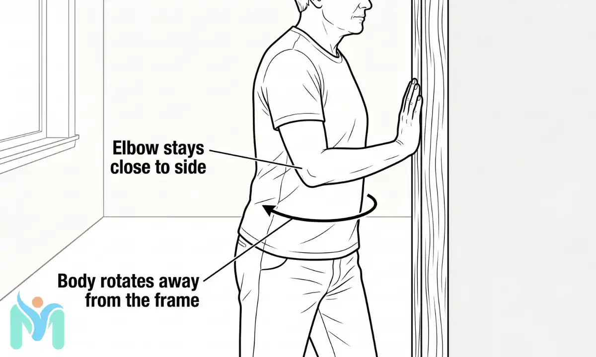
Purpose: External rotation is the most restricted movement in adhesive capsulitis, because the coracohumeral ligament and anterior joint capsule contract earliest in the condition. This stretch uses the door frame as a fixed reference point, allowing the body’s own rotation to progressively lengthen these structures without any active shoulder muscle effort working against the restriction.
Start position: Stand in a doorway or facing a wall. Bend your elbow to 90 degrees and keep it pressed gently against your side. Place your forearm and palm flat against the door frame or wall surface.
Movement: Keeping your elbow at your side, rotate your body slowly away from your forearm. The contact between your forearm and the frame creates the external rotation stretch at the front of the shoulder. Hold for 20 to 30 seconds, then return.
Dosage: 3 to 5 repetitions, holding 20 to 30 seconds each, once or twice daily.
Safety note: The elbow must stay close to your side throughout. Letting it drift forward or outward removes the specificity of the stretch for the coracohumeral ligament.
Stage 3 exercises: Resolution (Thawing) stage
As the shoulder enters Stage 3, movement is returning and the joint tolerates active exercises. The priority shifts from lengthening contracted tissue to rebuilding the active strength and motor control that were suppressed during months of restricted movement. The rotator cuff muscles, which have been inhibited throughout the earlier stages, need progressive retraining.
5. Active wall walk
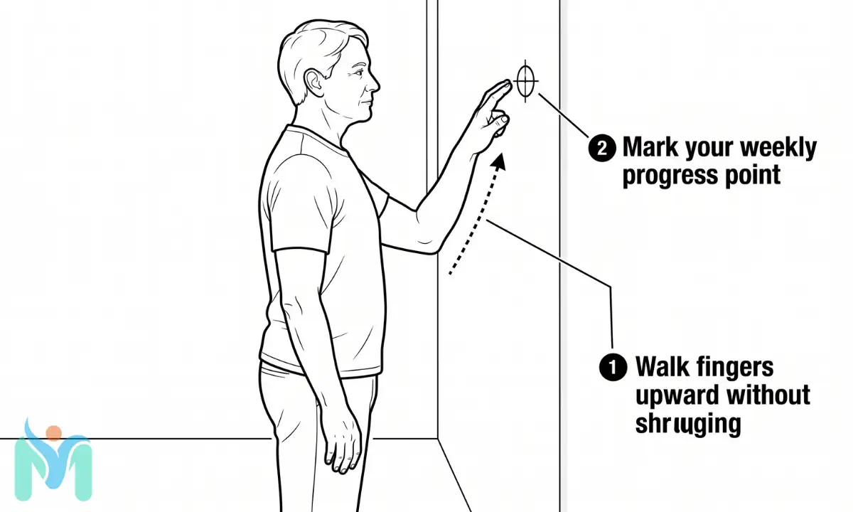
Purpose: The wall walk rebuilds active shoulder elevation progressively, using the wall surface as both a support and a weekly measurement reference. It retrains the supraspinatus and deltoid muscles to produce controlled overhead reach in small, trackable increments, making recovery visible and motivating at a stage when progress can otherwise feel slow.
Start position: Stand facing a wall, close enough to reach it easily. Place the fingertips of your affected arm on the wall surface at waist height.
Movement: Walk your fingers upward along the wall, step by step, reaching as high as you can without shrugging the shoulder or leaning sideways. Hold the highest point for 3 to 5 seconds. Walk fingers slowly back down. Mark the highest point reached after each session to track weekly progress.
Dosage: 10 repetitions, 2 sets, twice daily. Aim to increase height by 1 to 2 centimetres each week.
Safety note: Stop if you feel a sharp catching sensation at the top of the shoulder range. Increase height gradually each week rather than forcing range all at once.
6. Towel-assisted internal rotation reach
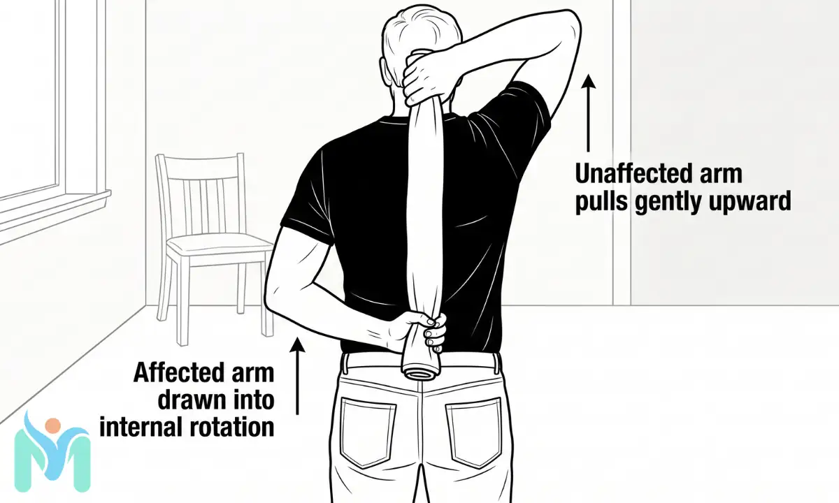
Purpose: Internal rotation behind the back (reaching the hand up the spine, as when fastening clothing) is one of the last movements to return in the thawing stage because the posterior inferior capsule and teres minor remain restricted longest. The towel transfers the mechanical effort to the unaffected arm, drawing the affected arm into the restricted range without requiring it to generate force against its own limitation.
Start position: Stand upright. Hold one end of a rolled towel in your affected hand, with that arm reaching behind your back to approximately hip or lower-back height. Reach your unaffected arm up and over the same shoulder to hold the other end of the towel from above.
Movement: Using your unaffected arm, pull the towel gently upward. This draws your affected hand progressively further up your back into internal rotation. Hold the furthest comfortable position for 15 to 20 seconds, then slowly release.
Dosage: 3 to 5 repetitions, holding 15 to 20 seconds each, twice daily.
Safety note: Pull gently and progressively. Do not jerk the towel. Release immediately if you feel pins and needles or numbness anywhere in the arm or hand.
For progression beyond these starting exercises, a physiotherapist can design the next phase of your programme based on the specific pattern of restriction remaining in your shoulder.
When exercises alone aren’t enough: what else can help?
Exercise is the foundation of frozen shoulder management, and it works alongside other options rather than in isolation. For people in Stage 1 whose pain is severe enough to limit exercise tolerance, a corticosteroid injection (a guided anti-inflammatory injection into the shoulder joint or surrounding space) can reduce pain in the short term. The 2011 CSP clinical practice guideline led by Hanchard and colleagues notes that injections provide meaningful short-term pain relief but do not alter the long-term course of the condition, which is why exercise remains the primary recovery tool throughout all stages.
Hydrodilation (arthrographic distension, where a saline solution is injected into the contracted joint capsule under pressure to stretch it from the inside) is used by some shoulder specialists during Stage 2 when physiotherapy alone has not produced sufficient progress after several months. Evidence for this approach is moderate rather than definitive, and it is generally considered when conservative management has stalled.
For a smaller group of patients who do not recover adequately after 9 to 12 months of consistent conservative management, manipulation under anaesthesia (MUA, in which the shoulder is moved through its range while the patient is sedated) or arthroscopic capsular release (a procedure in which the contracted joint capsule is cut surgically to restore joint volume) may be discussed with an orthopaedic consultant. These are later interventions, not first-line options.
Applying heat to the shoulder for 10 to 15 minutes before stretching exercises can help relax the capsular tissue and improve the quality of the stretch. Ice applied for 10 to 15 minutes after exercise may reduce post-exercise discomfort during the more reactive Stage 1 period.
When to expect real progress with frozen shoulder
Frozen shoulder exercises are not a quick fix, and the timeline is one of the harder realities to make peace with. The most-cited longitudinal study on natural outcomes, published by Reeves in 1975, found average resolution took approximately 30 months from symptom onset. Physiotherapy does not always shorten the total timeline, but it reduces the period of functional disability substantially, and that is what matters most for daily life.
Most people notice a meaningful reduction in night pain at around 6 weeks of consistent exercise. That shift is the first reliable sign the condition is responding. Full movement returns more slowly, and external rotation tends to recover last. The coracohumeral ligament, the structure that contracts earliest in frozen shoulder, is also the last to lengthen, which is why overhead reach and behind-back activities may feel restricted even after general shoulder comfort has improved considerably.
Recovery is non-linear. Most weeks bring small incremental progress interrupted by the occasional harder day, and a difficult morning does not undo the work of the weeks before it. Stay consistent with stage-appropriate frozen shoulder exercises, track which movements are returning, and work with a physiotherapist to progress the programme as your range increases. The shoulder that feels impossible to lift today will, with the right management, move freely again.
Consult your doctor or a qualified physiotherapist before starting any new exercise programme, especially if you have an existing injury or medical condition.
Frequently Asked Questions (FAQs)
The fastest recovery from frozen shoulder comes from consistent stage-appropriate physiotherapy exercises, combined with a corticosteroid injection in the early painful stage if pain is severe enough to limit exercise tolerance. According to the 2012 Chartered Society of Physiotherapy evidence-based guideline, no single treatment has been shown to be definitively superior in the long term, but early physiotherapy engagement combined with short-term pain management produces the best functional outcomes. Avoiding aggressive stretching during Stage 1 also protects against prolonged inflammation.
Stage 1 (Painful) is characterised by severe aching pain, often worst at night, with stiffness only beginning to develop. Stage 2 (Frozen) brings dramatic stiffness in all directions, particularly in external rotation, with less intense pain than Stage 1. Stage 3 (Thawing) sees movement gradually returning with settling discomfort. Frozen shoulder typically restricts external rotation more than abduction, and abduction more than internal rotation. A physiotherapist can confirm your stage and identify your specific pattern of restriction through a clinical assessment.
Whether to work through pain depends on the type of pain. A gentle pulling or mild ache during a stretch that fades within 30 minutes is normal, expected therapeutic discomfort. Sharp, sudden, or worsening pain during exercise, or pain that persists more than an hour after finishing, is a warning sign to reduce intensity and rest. In Stage 1, exercise within the pain-free range. In Stage 2, mild discomfort during sustained stretching is acceptable and expected as the contracted capsule is progressively lengthened.
In Stage 1, avoid forceful stretching, overhead pressing, or any exercise that produces pain lasting beyond the session. High-impact shoulder exercises and aggressive manual stretching are not appropriate during the inflammatory phase, as they can provoke the synovial lining and extend the painful stage. Across all stages, avoid any exercise causing sharp sudden pain or that produces increased swelling and warmth in the joint after completion. If unsure whether a specific exercise suits your stage, have it assessed by a physiotherapist before starting.
Both physiotherapy and corticosteroid injections have a role in frozen shoulder management, but they serve different purposes. Injections provide meaningful short-term pain relief, particularly in Stage 1, which can make exercise more tolerable. Physiotherapy exercise is the primary driver of long-term movement recovery. According to the 2011 CSP clinical practice guideline led by Hanchard and colleagues, injections do not change the long-term outcome of the condition – physiotherapy exercise is the treatment that drives movement recovery over time.
Recurrence in the same shoulder after full resolution is uncommon. However, research suggests frozen shoulder develops in the opposite shoulder in approximately 20 to 30% of people who experience it on one side, typically within 5 years of the first episode. The second episode follows the same clinical stages but does not always produce the same degree of stiffness or pain. Maintaining general shoulder mobility, managing underlying risk factors such as diabetes, and starting physiotherapy early if symptoms develop on the other side all reduce the functional impact of a second episode.
References
- Hanchard NC, Goodchild L, Thompson J, et al. “Evidence-based clinical guidelines for the diagnosis, assessment and physiotherapy management of contracted (frozen) shoulder.” Physiotherapy. 2012 Jun;98(2):117-20. DOI: 10.1016/j.physio.2012.01.001. PMID: 22507361. URL: https://pubmed.ncbi.nlm.nih.gov/22507361/. Evidence Level: 1.
- Kelley MJ, Shafer AP, McCormick TJ, et al. “Shoulder pain and mobility deficits: adhesive capsulitis. Clinical practice guidelines linked to the International Classification of Functioning, Disability, and Health from the Orthopaedic Section of the American Physical Therapy Association.” J Orthop Sports Phys Ther. 2013;43(5):A1-31. DOI: 10.2519/jospt.2013.0302. PMID: 23636125. URL: https://pubmed.ncbi.nlm.nih.gov/23636125/. Evidence Level: 1.
- Page MJ, Green S, Kramer S, Johnston RV, McBain B, Buchbinder R. “Manual therapy and exercise for adhesive capsulitis (frozen shoulder).” Cochrane Database Syst Rev. 2014;(8):CD011275. DOI: 10.1002/14651858.CD011275. URL: https://www.cochranelibrary.com/cdsr/doi/10.1002/14651858.CD011275/full. Evidence Level: 2.
- Dias R, Cutts S, Massoud S. “Frozen shoulder.” BMJ. 2005;331(7530):1453-1456. DOI: 10.1136/bmj.331.7530.1453. PMID: 16356211. URL: https://pubmed.ncbi.nlm.nih.gov/16356211/. Evidence Level: 2.
- Neviaser AS, Hannafin JA. “Adhesive capsulitis: a review of current treatment.” Am J Sports Med. 2010;38(11):2346-2356. DOI: 10.1177/0363546510374806. PMID: 20668229. URL: https://pubmed.ncbi.nlm.nih.gov/20668229/. Evidence Level: 2.
- Zreik NH, Malik RA, Charalambous CP. “Adhesive capsulitis of the shoulder and diabetes: a meta-analysis of prevalence.” Muscles Ligaments Tendons J. 2016;6(1):26-34. PMID: 27331029. URL: https://pubmed.ncbi.nlm.nih.gov/27331029/. Evidence Level: 2.
- Reeves B. “The natural history of the frozen shoulder syndrome.” Scand J Rheumatol. 1975;4(4):193-196. PMID: 1181428. URL: https://pubmed.ncbi.nlm.nih.gov/1181428/. Evidence Level: 5.
- NHS. “Frozen shoulder.” NHS Conditions. URL: https://www.nhs.uk/conditions/frozen-shoulder/ (2022). Evidence Level: 7.