


Staring at a pair of electrode pads and an unhelpfully vague placement diagram is the starting point for most people learning to use TENS. Whether you are a physiotherapy student encountering electrotherapy for the first time or a patient who has been given a TENS unit for back pain, the instruction “place over the area of pain” leaves most of the important clinical decisions unanswered.
TENS electrode placement determines which nerve fibres the current actually reaches and, by extension, whether the stimulation has any genuine effect on pain. Reaching the right fibres engages a specific neurophysiological mechanism. Placing the pads without that logic produces only a tingling sensation with no therapeutic target.
Why TENS electrode placement actually matters
Transcutaneous electrical nerve stimulation (TENS) works by delivering a low-level electrical current through adhesive electrode pads placed on the skin. Where those pads sit determines which nerve fibres the current reaches and whether pain signals are interrupted at the level of the spinal cord. Correct TENS electrode placement targets the specific neural pathways responsible for the pain by positioning pads over or near the painful area, along a peripheral nerve trunk, or alongside the spine at the corresponding spinal segment.
Research published in Physical Therapy by Sluka and colleagues in 2013 noted that inconsistent outcomes in TENS clinical trials are partly explained by poor treatment fidelity, which includes inadequate electrode placement. The electrode positioning decisions made before you switch the machine on may determine the outcome more than the device settings themselves.
Pain is processed at multiple levels: at the peripheral tissue where damage is detected, at the dorsal horn of the spinal cord where incoming signals are modulated, and at higher brain centres where the experience of pain is generated. Electrode placement needs to account for where along that pathway the intervention is most likely to be effective, and that depends on both the condition being treated and the mode of TENS being applied.
How TENS works, and why it changes where you place the pads
There are two clinically distinct modes of TENS, and they work through completely different neurological mechanisms. Understanding that distinction is what makes electrode placement logical rather than arbitrary.
Conventional TENS uses a high frequency, typically 80 to 150 Hz, at a low intensity that produces a comfortable tingling sensation called paresthesia. This level of stimulation preferentially activates large-diameter Aβ fibres, which carry information about light touch and pressure. In their foundational 1965 paper in Science, Melzack and Wall described the gate control theory of pain: Aβ fibre activity activates inhibitory interneurons in the dorsal horn of the spinal cord, suppressing the transmission of pain signals from smaller-diameter C fibres and Aδ fibres. The tingling of conventional TENS is, in effect, activating your nervous system’s own pain suppression mechanism.
For conventional TENS, electrode placement should produce tingling that covers or surrounds the painful area. The current needs to reach the Aβ fibres serving that specific skin region, which requires the electrodes to sit so the current path runs through the painful territory.
Acupuncture-like TENS (AL-TENS) operates at a low frequency of 1 to 4 Hz at an intensity high enough to produce a visible muscle twitch. A study by Sjölund and Eriksson, published in Brain Research in 1979, demonstrated that this type of stimulation triggers the release of endogenous opioids, including β-endorphins, from the central nervous system. This analgesic effect was reversed by naloxone, an opioid antagonist, confirming that the mechanism relies on the body’s own opioid system. For AL-TENS, electrodes target motor points or acupuncture points within the myotome (the muscle group sharing the same spinal segment as the pain) rather than directly over the pain site.
Because conventional TENS and AL-TENS work through entirely different mechanisms, they require different electrode placement strategies, and most home users apply only one type, usually without knowing which one.
A review by Sluka and Walsh, published in the Journal of Pain in 2003, confirmed that these distinct mechanisms require distinct placement protocols to produce their intended clinical effects.
The 4 placement strategies physiotherapists use
TENS electrode placement in clinical physiotherapy practice follows four main strategies. Each targets a different point along the pain processing pathway, and the right choice depends on the condition, the mode of TENS, and where symptoms are concentrated.
- Over the site of pain: Both electrodes are placed on or immediately around the painful area. This is the standard approach for conventional TENS and works well for localised musculoskeletal pain where the skin over the pain site shares the same nerve supply as the painful structure beneath. The paresthesia produced should cover the area of discomfort, confirming correct targeting.
- Paravertebral placement: Electrodes are positioned on either side of the spine at the vertebral level corresponding to the pain’s spinal segment, targeting the dorsal root ganglion and spinal nerve at the point where pain signals enter the spinal cord. A review by Vance and colleagues in Pain Management (2014) describes paravertebral positioning as a valid clinical strategy for pain with a clear dermatomal or segmental distribution.
- Over a peripheral nerve trunk: One or both electrodes are placed directly over an accessible peripheral nerve rather than over the pain site itself, such as over the common peroneal nerve at the fibular head for foot pain, or over the median nerve at the wrist for hand symptoms. This provides direct stimulation of the nerve supplying the painful area.
- At motor points or acupuncture points: Used primarily with AL-TENS, this strategy targets motor points within the painful myotome to produce a muscle twitch and drive endogenous opioid release. Motor point mapping requires clinical training and is better suited to supervised physiotherapy than home use.
TENS electrode placement by condition: a practical guide
The placements below reflect common clinical protocols based on the neuroanatomy of each condition. These are starting points, not fixed prescriptions. Exact placement may need adjusting based on where your symptoms are most concentrated and how the tingling sensation actually spreads once the unit is running.
Lower back pain
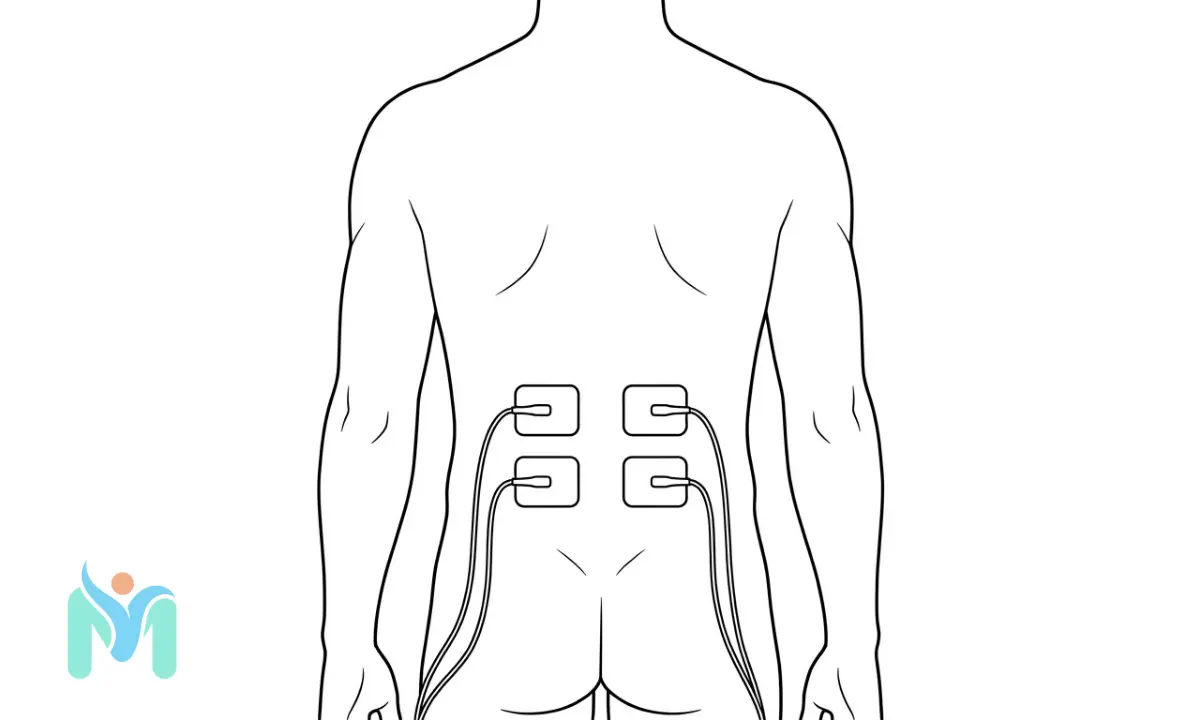
Place two or four electrodes paravertebrally at the lumbar spinal level corresponding to the pain, typically between L1 and L5. Position each pad approximately 2 to 3 cm from the midline of the spine on each side, over the paraspinal muscles rather than directly on the vertebral spinous processes. Increase intensity until you feel comfortable tingling covering the painful area without any sharp or burning sensation.
A 2008 Cochrane review by Khadilkar and colleagues, comparing TENS against placebo for chronic low back pain, found evidence of short-term pain reduction, with paravertebral placement being the standard protocol across the included trials. If pain radiates into the buttock without extending down the leg, placing the lower pair of electrodes over the gluteal region alongside the paravertebral pair can extend the coverage area. Stop the session and seek advice if you notice any new shooting pain down the leg during treatment.
Sciatica and leg pain
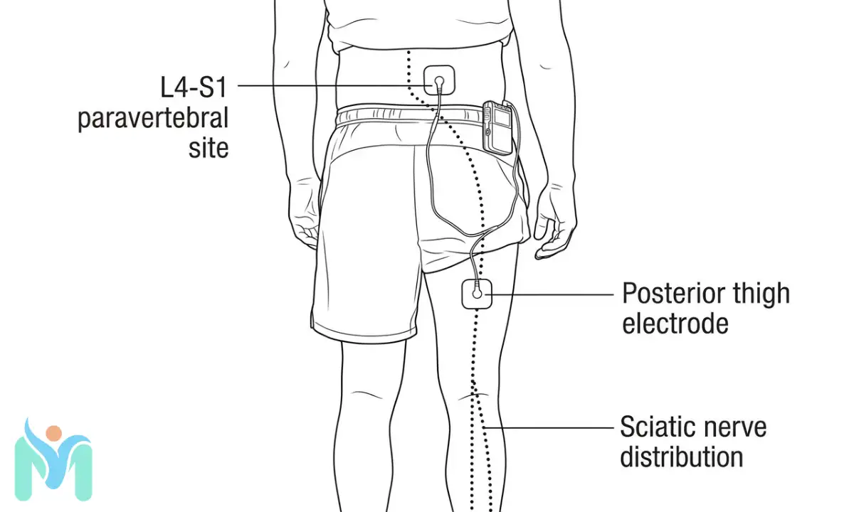
The sciatic nerve arises from the L4, L5, and S1 to S3 nerve roots and travels through the buttock and along the posterior leg. A combination placement that targets both the nerve’s spinal origin and its peripheral distribution produces better coverage than placing electrodes at a single point. Position one electrode paravertebrally at L4 to S1 and a second along the path of radiating pain in the leg: the posterior thigh, the calf, or the ankle, depending on where symptoms are strongest.
If reduced skin sensation in the foot or leg is part of your presentation, avoid placing electrodes over areas where you cannot reliably detect whether the intensity is too high.
Knee pain
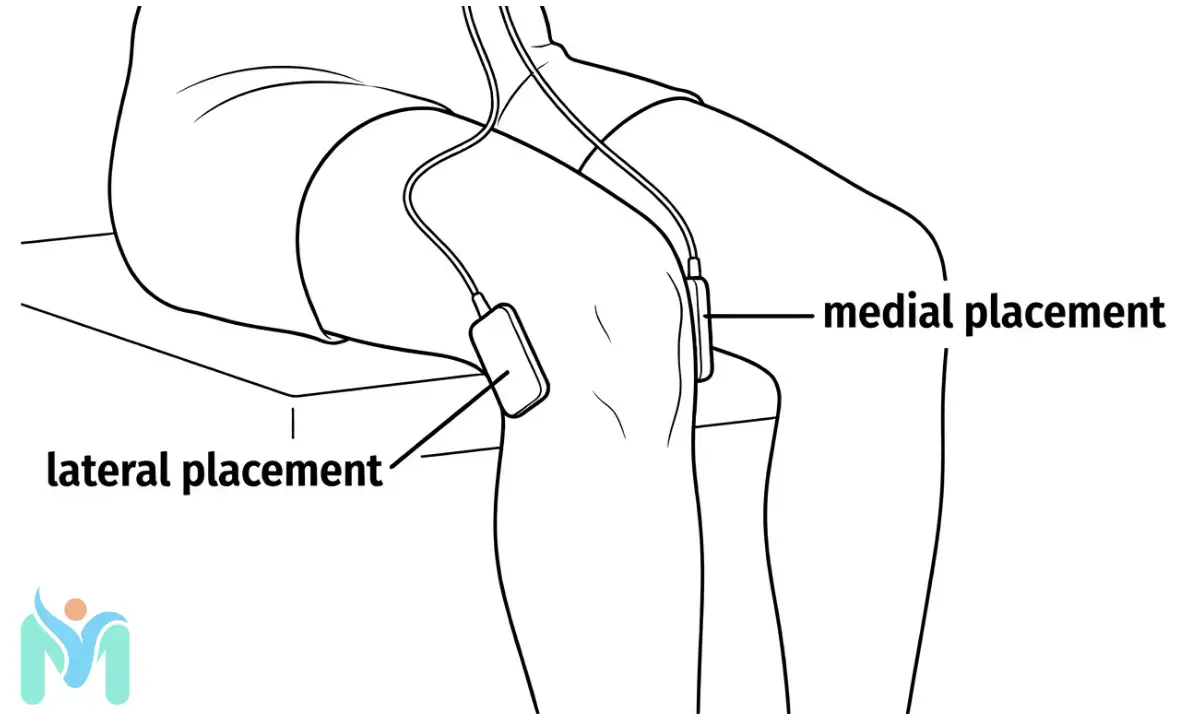
For knee pain, including osteoarthritis, place electrodes medially and laterally to the joint line at the same horizontal level, flanking the knee joint. This cross-joint configuration allows current to pass through the joint space. One electrode just above the patella and one just below covers the anterior knee and infrapatellar region as an alternative.
A 2009 Cochrane review by Rutjes and colleagues examined electrical stimulation for knee osteoarthritis and found a modest but measurable reduction in pain intensity, supporting TENS as an adjunct in managing this condition. Remove the electrodes immediately if you feel any burning sensation under the pad.
Shoulder pain
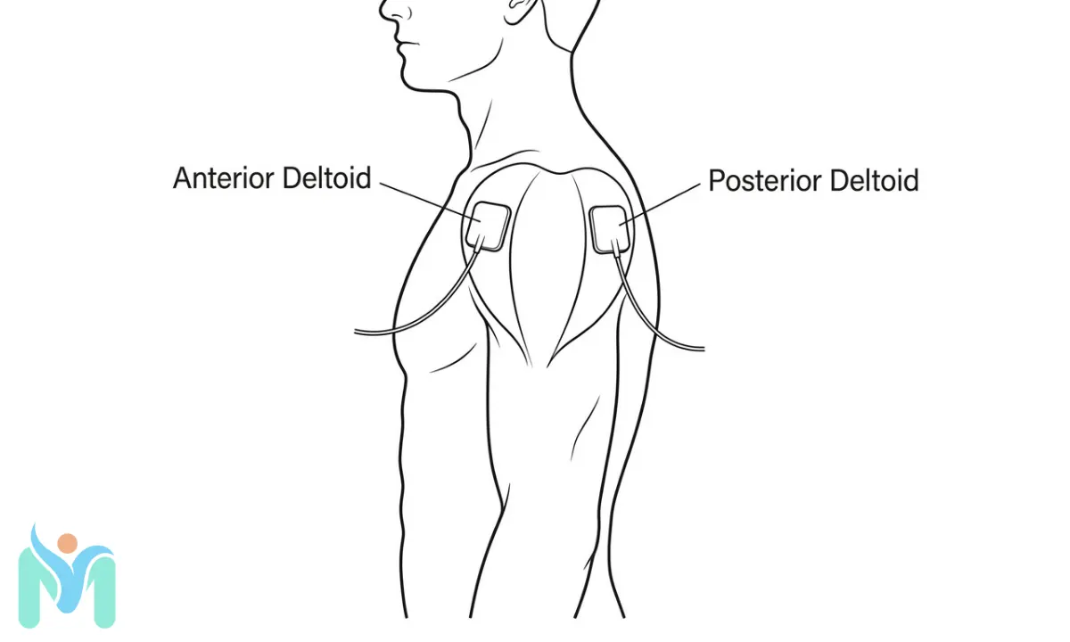
Position one electrode over the anterior deltoid and one over the posterior deltoid, flanking the shoulder joint. This passes current through the joint. For pain from the supraspinatus or upper rotator cuff, moving the upper electrode to the lower trapezius muscle just above the shoulder targets the C4 to C5 dermatomal level that supplies the shoulder region.
Keep all electrodes well clear of the anterior neck. The area over the carotid sinus, located at the pulse point on either side of the neck just below the jaw, is an absolute contraindication covered in the safety section below.
Neck pain
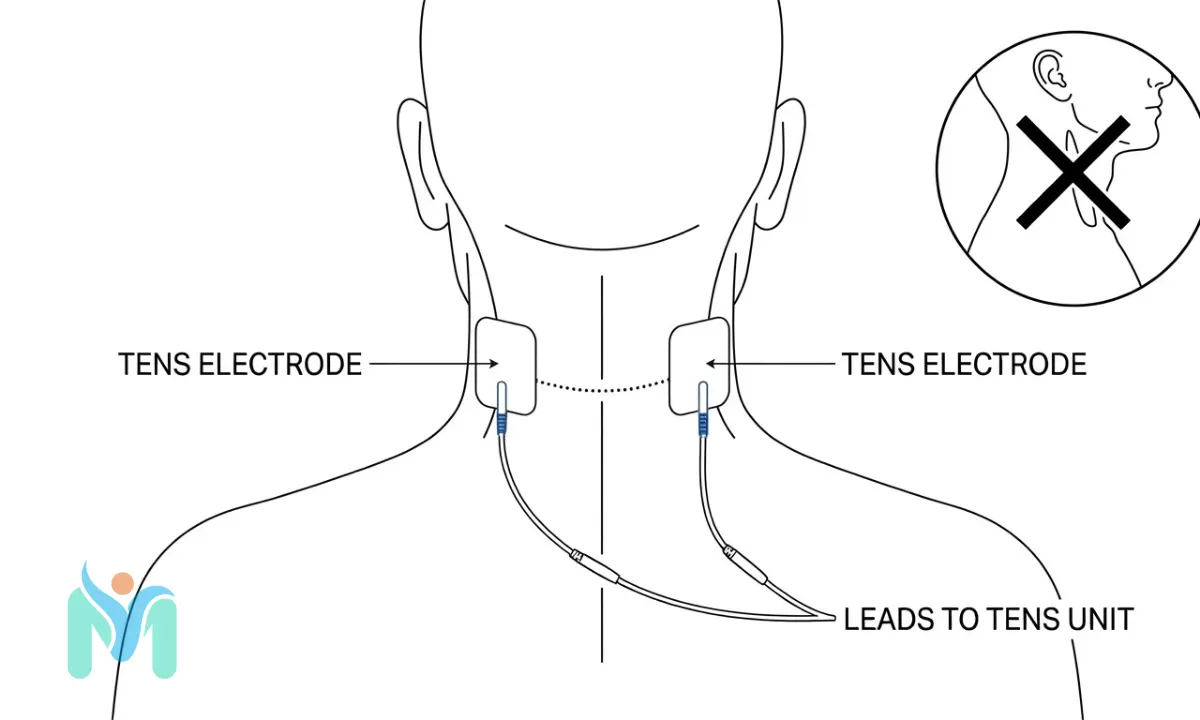
For cervical pain, place electrodes bilaterally on the paraspinal muscles of the posterior neck at approximately C4 to C7 level, sitting on the muscles flanking the vertebral column rather than directly on the spinous processes. Both electrodes must remain on the posterior neck throughout the session.
Never place TENS electrodes over the front of the neck, the throat, or the area of the carotid pulse. Stimulation of the carotid sinus triggers a baroreceptor reflex that can cause sudden hypotension and cardiac arrhythmia. This is an absolute contraindication, not a precaution to weigh against potential benefit.
How far apart should TENS electrode pads be?
Electrode spacing determines the depth of the current path through tissue. Electrical current follows the path of least resistance between the two pads. When pads are close together, that path is shallow, stimulating superficial nerves and skin receptors. When pads are further apart, the current takes a deeper arc to travel between them, reaching deeper muscles, joint capsules, and nerve roots.
Chesterton and colleagues, in a 2002 study published in Pain, demonstrated that placement variables, including electrode positioning relative to the target nerve, produce measurable differences in pain threshold outcomes in healthy subjects. Spacing decisions are part of how TENS is dosed, not peripheral details.
The minimum safe distance between electrode edges is approximately 2 to 3 cm. At shorter distances, current bridges across the skin surface rather than penetrating the tissue. For large or deep structures such as the lumbar paraspinal muscles, separating electrodes by 8 to 15 cm allows the current to reach deeper tissue layers.
| Target area | Approximate electrode separation | Notes |
|---|---|---|
| Small or superficial joints (wrist, ankle, fingers) | 3 to 5 cm | Current stays superficial; suitable for stimulating surface nerves |
| Knee joint and shoulder joint | 6 to 10 cm | Cross-joint or flanking placement to reach intra-articular structures |
| Lumbar spine and deep paraspinal structures | 8 to 15 cm | Wider separation allows a deeper current arc through paraspinal muscles |
| Bilateral paravertebral placement | Width of the spine between the two pads | One pad each side: current crosses through the spinal segment area |
Before applying electrodes, clean the skin with warm water and dry it thoroughly. Remove any lotion, oil, or perspiration from the area, as these increase skin impedance and reduce current transmission efficiency. Inspect the skin for redness, cuts, or irritation before each session, and check the skin again after removing the pads. If redness under an electrode does not resolve within 30 minutes, adjust the placement site at the next session and seek advice if the redness persists or worsens.
Where should you never place TENS electrodes?
Certain placements carry genuine clinical risk and are contraindicated regardless of pain intensity or location. According to NHS guidance on TENS, the following are firm restrictions.
- Over or near the heart: Electrode placement that passes current across the thorax, front to back or side to side over the chest, can interfere with cardiac rhythm.
- Over an implanted cardiac pacemaker, spinal cord stimulator, or cochlear implant: Electrical current can disrupt device function and endanger the wearer. This applies to the area over both the device body and its electrode leads.
- Over the front of the neck or carotid sinus: Stimulation at the carotid sinus triggers a baroreceptor reflex causing an abrupt drop in blood pressure and potential cardiac arrhythmia.
- Transcerebrally, across the head or skull: Passing current through brain tissue is not safe at home TENS intensities for untrained users.
- Over the eyes: Risk of damage to the retina and optic nerve.
- Over the abdomen or lower back during pregnancy: The risk of uterine stimulation means this placement requires obstetric supervision. NHS guidance states that TENS should not be used during pregnancy without medical clearance. TENS is used for labour pain in supervised clinical settings under specific protocols, but home use during pregnancy is a separate matter that requires professional advice first.
- Over broken, infected, or irritated skin: Current through damaged skin increases the risk of a skin burn and may worsen existing tissue damage.
- Over a known deep vein thrombosis: Stimulation over an active DVT carries a theoretical risk of clot dislodgement.
People with epilepsy, impaired skin sensation, or cognitive impairment should seek clinical guidance before using TENS, as they may be unable to recognise an adverse response to placement or to intensity that is too high.
What the evidence really says about TENS
TENS has been studied extensively, and an honest reading of that evidence requires acknowledging both where the support is clear and where it is limited. Many patients are surprised to find that results vary so much between users. The explanation is usually found in how the therapy was applied rather than in whether it works at all.
A 2015 Cochrane review by Johnson and colleagues, covering TENS for both acute and chronic pain, concluded that while TENS shows promise across multiple conditions, the overall evidence quality is low, with many trials using inadequate dosing and inconsistent electrode placement. Bennett and colleagues, in a 2011 analysis published in Pain, found that low treatment fidelity, including suboptimal electrode positioning, was a consistent problem across TENS trials. Many negative findings in the TENS research literature may reflect inadequate application of the therapy rather than a failure of the underlying mechanism.
For specific conditions, the picture is more defined. The 2009 Cochrane review by Rutjes and colleagues found a modest but measurable reduction in pain for knee osteoarthritis. The 2008 Cochrane review by Khadilkar and colleagues found evidence of short-term pain reduction for chronic low back pain. These findings support clinical use of TENS as an adjunct rather than as a replacement for broader physiotherapy management.
The clearest boundary in the evidence comes from NICE’s 2021 guideline on chronic primary pain (NG193), which recommends against offering TENS for this specific diagnostic category due to insufficient evidence. This guidance applies to conditions such as chronic widespread pain and chronic primary musculoskeletal pain. It does not preclude TENS use for osteoarthritis, post-surgical pain, labour pain, or acute musculoskeletal conditions, where clinical use continues with reasonable evidential support.
Making TENS work for you
The research points consistently to one practical conclusion: how TENS is applied determines whether it works. The clinical failures in TENS research are not all failures of the intervention; many reflect failures of technique, particularly electrode placement that did not reach the target nerve fibres. That is a solvable problem.
The TENS electrode placement protocols in this guide are based on the neuroanatomy of each pain distribution and the clinical protocols used in physiotherapy practice. At MystPhysio.com, all rehabilitation content is reviewed by qualified physiotherapists and updated in line with current clinical guidelines. Starting from a placement grounded in dermatomal anatomy and verified clinical practice gives you a genuinely stronger foundation than placing pads wherever feels most convenient.
For people managing chronic pain, TENS tends to be most effective as part of a broader self-management approach that includes graded activity, exercise, and physiotherapy guidance rather than as a standalone solution. If pain does not respond to TENS after several sessions using correct placement, that is clinically useful information. It may indicate the pain source lies beyond what electrode-based stimulation can reach, which points toward a different assessment or a different intervention.
TENS electrode placement is not a fixed procedure. It is a clinical reasoning process, and understanding the mechanism behind each strategy is what allows you to adapt it when your pain changes, when you switch between TENS modes, or when you move into a different stage of recovery.
Consult your doctor or a qualified physiotherapist before starting any new exercise programme, especially if you have an existing injury or medical condition.
Frequently Asked Questions (FAQs)
In most cases, TENS can be used safely over an area containing metal implants such as joint replacements or surgical plates. Metal implants do not meaningfully alter the current path in a way that causes harm at standard TENS intensities. However, some manufacturers advise against placement directly over the implant, so check your implant documentation first. Spinal cord stimulators and cardiac pacemakers are a separate matter and remain absolute contraindications for TENS at any intensity.
For lower back pain, place electrodes paravertebrally on either side of the lumbar spine at the level corresponding to your pain, typically between L1 and L5. Position each pad approximately 2 to 3 cm from the centre of the spine, over the paraspinal muscles rather than directly on the vertebral bone. Increase intensity gradually until you feel tingling covering the painful area. A physiotherapist can map the correct level for your specific pain pattern if you are unsure.
Most TENS sessions last 20 to 60 minutes. There is no firm upper limit in clinical guidelines, but using TENS continuously at the same setting for several hours is not recommended without clinical supervision, as the nervous system can adapt and the analgesic effect may diminish. NHS guidance recommends using TENS as needed while monitoring skin response. Taking short breaks between sessions or gradually adjusting intensity can help maintain effectiveness over time.
Skin irritation from TENS electrodes is usually caused by a reaction to the electrode adhesive, prolonged use over the same patch of skin, or intensity set too high. To reduce risk, clean the skin before each session, rotate placement sites slightly between sessions, and inspect the skin after removing pads. Any redness, blistering, or burning sensation that persists more than 30 minutes after electrode removal should prompt you to stop and seek clinical advice before the next session.
TENS should not be used over the abdomen or lower back during pregnancy without medical supervision, due to the risk of uterine stimulation. TENS is used for labour pain management in supervised obstetric settings following specific clinical protocols, but home use during pregnancy requires medical clearance. According to NHS guidance on TENS, it is not recommended during pregnancy without professional advice. Speak with your midwife or obstetrician before using TENS if you are pregnant.
Do not place TENS electrodes directly over the vertebral spinous processes, the bony prominences running along the midline of your back. The correct placement target for spinal pain is the paraspinal muscles, which sit 2 to 3 cm lateral to the midline on each side. These muscles provide the surface through which the current reaches the dorsal root ganglia and spinal nerves that are the actual therapeutic targets for back and neck pain. Placement on bone reduces stimulation efficiency.
If pain relief from TENS diminishes shortly after the session starts, this typically indicates accommodation: the nervous system adapts to constant stimulation at a fixed frequency and reduces its inhibitory response. To address this, gradually increase the intensity to re-establish the tingling sensation, or switch to a modulated or burst mode if your device offers one. Adjusting the electrode position slightly can also re-engage the sensory response. Accommodation is more common with conventional TENS at fixed parameters than with AL-TENS.
References
- Sluka KA, Bjordal JM, Marchand S, Rakel BA. “What Makes Transcutaneous Electrical Nerve Stimulation Work? Making Sense of the Inconsistent Results in the Clinical Literature.” Phys Ther. 2013;93(10):1397-402. DOI: 10.2522/ptj.20120281. PMID: 23813082. URL: https://pubmed.ncbi.nlm.nih.gov/23813082/. Evidence Level: 5.
- Melzack R, Wall PD. “Pain mechanisms: a new theory.” Science. 1965;150(3699):971-9. DOI: 10.1126/science.150.3699.971. PMID: 5320816. URL: https://pubmed.ncbi.nlm.nih.gov/5320816/. Evidence Level: 4.
- Sluka KA, Walsh D. “Transcutaneous electrical nerve stimulation: basic science mechanisms and clinical effectiveness.” J Pain. 2003;4(3):109-21. DOI: 10.1054/jpai.2003.434. PMID: 14622708. URL: https://pubmed.ncbi.nlm.nih.gov/14622708/. Evidence Level: 5.
- Sjölund BH, Eriksson MB. “The influence of naloxone on analgesia produced by peripheral conditioning stimulation.” Brain Res. 1979;173(2):295-301. PMID: 455454. URL: https://pubmed.ncbi.nlm.nih.gov/455454/. Evidence Level: 4.
- Vance CGT, Dailey DL, Rakel BA, Sluka KA. “Using TENS for pain control: the state of the evidence.” Pain Manag. 2014;4(3):197-209. DOI: 10.2217/pmt.14.13. PMID: 24953072. URL: https://pubmed.ncbi.nlm.nih.gov/24953072/. Evidence Level: 5.
- Khadilkar A, Odebiyi DO, Brosseau L, Wells GA. “Transcutaneous electrical nerve stimulation (TENS) versus placebo for chronic low-back pain.” Cochrane Database Syst Rev. 2008;(4):CD003008. DOI: 10.1002/14651858.CD003008.pub3. PMID: 18843638. URL: https://pubmed.ncbi.nlm.nih.gov/18843638/. Evidence Level: 2.
- Rutjes AW, Nüesch E, Sterchi R, Kalichman L, Hendriks E, Osiri M, Brosseau L, Reichenbach S, Jüni P. “Transcutaneous electrostimulation for osteoarthritis of the knee.” Cochrane Database Syst Rev. 2009;(4):CD002823. DOI: 10.1002/14651858.CD002823.pub2. PMID: 19821296. URL: https://pubmed.ncbi.nlm.nih.gov/19821296/. Evidence Level: 2.
- Chesterton LS, Barlas P, Foster NE, Lundeberg T, Wright CC, Baxter GD. “Sensory stimulation (TENS): effects of parameter manipulation on mechanical pain thresholds in healthy human subjects.” Pain. 2002;99(1-2):253-62. DOI: 10.1016/S0304-3959(02)00118-1. PMID: 12237205. URL: https://pubmed.ncbi.nlm.nih.gov/12237205/. Evidence Level: 4.
- Johnson MI, Paley CA, Howe TE, Sluka KA. “Transcutaneous electrical nerve stimulation for acute and chronic pain.” Cochrane Database Syst Rev. 2015;(6):CD009584. DOI: 10.1002/14651858.CD009584.pub2. PMID: 26121650. URL: https://pubmed.ncbi.nlm.nih.gov/26121650/. Evidence Level: 2.
- Bennett MI, Hughes N, Johnson MI. “Methodological quality in randomised controlled trials of transcutaneous electric nerve stimulation for pain: low fidelity may explain negative findings.” Pain. 2011;152(6):1226-32. DOI: 10.1016/j.pain.2011.01.024. PMID: 21435786. URL: https://pubmed.ncbi.nlm.nih.gov/21435786/. Evidence Level: 5.
- NHS. “Transcutaneous electrical nerve stimulation (TENS).” NHS Conditions. URL: https://www.nhs.uk/conditions/transcutaneous-electrical-nerve-stimulation-tens/ (accessed 2024). Evidence Level: 7.
- NICE. “Chronic pain (primary and secondary) in over 16s: assessment of all chronic pain and management of chronic primary pain.” NICE guideline NG193. 2021. URL: https://www.nice.org.uk/guidance/ng193. Evidence Level: 1.