

One of the most common questions I hear in the clinic goes something like this: “My shoulder doesn’t go as far as my other one, is something wrong?” It’s a reasonable concern. But the honest answer is that “normal” joint movement is far more of a range than a single number. It shifts with age, sex, body type, activity level, and even which side of your body you’re measuring.
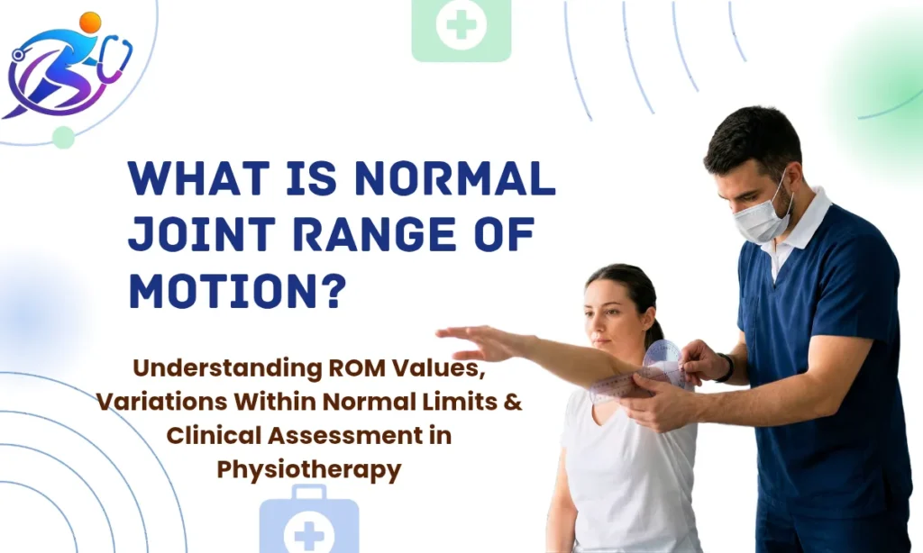
Range of motion, or ROM, refers to the full arc of movement a joint can travel through. Physiotherapists measure it routinely, using a tool called a goniometer (a hinged, protractor-like device) to record angles in degrees. These measurements guide clinical decisions: whether a joint is healing after injury, whether movement is restricted by pain or stiffness, or whether someone is progressing through rehabilitation.
This article covers what the research says about normal ROM values for major joints, what “within normal limits” actually means clinically, and why two people with perfectly healthy joints may move quite differently, and both be entirely fine.
What Is Joint Range of Motion?
Range of motion describes how far a joint can move in a specific direction. Every major joint in the body has defined movement planes, bending and straightening, rotating inward and outward, moving side to side.
There are two types:
- Active ROM (AROM): The movement you produce yourself using your own muscles.
- Passive ROM (PROM): The movement a clinician (or external force) produces for you, while your muscles are relaxed.
Passive ROM is usually slightly greater than active ROM. That gap between the two is clinically useful. A large difference can indicate muscle weakness, pain inhibition, or a neurological issue rather than a structural joint problem.
How Is ROM Measured?
The standard clinical tool is the goniometer. The clinician aligns the instrument’s axis over the joint and measures the angle of movement. Inclinometers and digital apps are also used in modern practice, often with better reproducibility for spinal measurements.
The American Academy of Orthopaedic Surgeons (AAOS) published widely used normative reference values. These form the baseline most clinicians compare against, though they are population averages, not absolute thresholds.
Normal ROM Values for Major Joints
Below are commonly cited normative values from AAOS guidelines and supporting literature. These are adult reference ranges under standard conditions.
Shoulder
The shoulder is the most mobile joint in the body. It trades stability for that mobility.
- Flexion (raising arm forward): 0-180°
- Extension (moving arm backward): 0-60°
- Abduction (raising arm to the side): 0-180°
- Internal rotation: 0-70°
- External rotation: 0-90°
In practice, I often find that overhead athletes, swimmers, tennis players, volleyball players, sit well above these upper values in external rotation. That isn’t abnormal. It reflects adaptation.
Elbow
- Flexion: 0-150°
- Extension: 0° (neutral straight position)
- Pronation (palm down rotation): 0-80°
- Supination (palm up rotation): 0-80°
Women tend to have a few degrees of natural hyperextension at the elbow, often up to 5-15° beyond neutral. This is a normal anatomical variant, not a sign of joint laxity or instability.
Wrist
- Flexion: 0-80°
- Extension: 0-70°
- Radial deviation (toward thumb): 0-20°
- Ulnar deviation (toward little finger): 0-30°
Hip
- Flexion: 0-120° (with knee bent); up to 90° with knee straight
- Extension: 0-30°
- Abduction (leg out to side): 0-45°
- Adduction (leg across midline): 0-30°
- Internal rotation: 0-45°
- External rotation: 0-45°
Hip ROM is highly age-dependent. Studies show a measurable decline in hip internal rotation and flexion beginning in the fourth decade of life. That’s not pathology, it’s a normal aging process, partly driven by changes in joint capsule elasticity and muscle length.
Knee
- Flexion: 0-135°
- Extension: 0° (full straight)
- Hyperextension: Up to 10° may be present in naturally lax individuals
Ankle and Foot
- Dorsiflexion (toes toward shin): 0-20°
- Plantarflexion (pointing foot down): 0-50°
- Inversion: 0-35°
- Eversion: 0-15°
Restricted ankle dorsiflexion, below 15°, is one of the more clinically significant findings in lower limb injury assessment. It has been linked to increased knee stress during squatting movements and a higher rate of ankle sprains.
Cervical Spine (Neck)
- Flexion: 0-45°
- Extension: 0-45°
- Lateral flexion (ear to shoulder): 0-45° each side
- Rotation: 0-60-80° each side
Lumbar Spine (Lower Back)
- Flexion: 0-60°
- Extension: 0-25°
- Lateral flexion: 0-25° each side
Spinal measurements are notably more variable and harder to standardise than limb joints. Inclinometry is more reliable than goniometry here.
RELATED: The 80/20 Rule: Best Home Balance Exercises for Seniors
Variations Within Normal Limits: Why People Differ
Age-Related Changes
ROM generally decreases with age. A systematic review published in the Journal of Aging and Physical Activity confirmed progressive loss of hip, knee, and ankle mobility across the lifespan, with the steepest decline typically occurring after age 55. This is largely due to reduced water content in joint cartilage, capsule thickening, and loss of muscle extensibility. It does not automatically indicate disease.
Sex Differences
Females tend to have greater ROM than males across most joints, particularly at the hip and spine. Hormonal influences, particularly the role of relaxin, contribute to greater ligamentous laxity. This difference is most pronounced in the reproductive years.
Hypermobility
Some people have generalised joint hypermobility, joints that move beyond the standard upper limits in multiple regions. This affects an estimated 10-20% of the population to some degree. The Beighton Score is a quick clinical screening tool for this.
Hypermobility is not always a problem. Many hypermobile individuals are asymptomatic. However, a subset develops chronic joint pain, fatigue, and proprioceptive difficulties, a condition now recognised as Hypermobile Ehlers-Danlos Syndrome (hEDS) or Hypermobility Spectrum Disorder (HSD). Both require specialist assessment.
Dominance and Side-to-Side Asymmetry
Minor asymmetry between sides is expected. Dominant-side shoulders in throwing athletes often show increased external rotation and decreased internal rotation compared to the non-dominant side, a well-documented adaptation called Glenohumeral Internal Rotation Deficit (GIRD). In isolation, small side-to-side differences are not clinically alarming. It is significant asymmetry, typically more than 10-15°, that warrants closer investigation.
Activity Level and Training History
Yoga practitioners and gymnasts often demonstrate ROM values well beyond population norms. Conversely, people with sedentary occupations may show restricted hip flexion or thoracic mobility, not from pathology, but from prolonged static postures and underuse.
When Restricted ROM Becomes a Clinical Concern
Not all restriction is pathological, but certain patterns are. Physiotherapists look at:
- End-feel – what the joint “feels like” at its limit. A firm elastic stop is different from a hard bony block or an empty (pain-limited) stop.
- Pattern of restriction – certain capsular patterns (predictable combinations of movement loss) suggest specific diagnoses. For example, the capsular pattern of the shoulder involves greatest loss of external rotation, followed by abduction, then internal rotation.
- Functional impact – a restriction matters most when it prevents daily activities: reaching overhead, walking without a limp, turning the head to drive safely.
- Symptom association – is the restriction painful throughout the range, only at the limit, or not painful at all?
ROM measurement is never interpreted in isolation. A good clinician reads the numbers alongside the clinical story.
The Role of ROM Assessment in Physiotherapy Practice
ROM assessment serves several purposes in physiotherapy:
- Establishing a baseline at first assessment
- Monitoring progress during rehabilitation
- Identifying asymmetries that may predict injury risk
- Guiding exercise prescription – knowing the available range helps set safe parameters for strength training and mobility work
- Communicating outcomes with the broader healthcare team in a standardised, measurable way
Reliable ROM measurement requires consistent technique. Small variations in patient positioning, stabilisation, and goniometer placement can meaningfully affect recorded angles. Inter-rater reliability (consistency between different clinicians) is generally better at the hip and knee than at the shoulder or spine.
Frequently Asked Questions (FAQs)
Normal knee flexion is 0-135°, and full extension is 0° (straight). Some people naturally reach 135-145° without any stretching. Hyperextension up to about 10° can be a normal variant, particularly in women. If you cannot fully straighten your knee after an injury, that warrants assessment.
Yes, in many cases. ROM restrictions caused by muscle tightness, scar tissue, post-surgical stiffness, or disuse respond well to targeted physiotherapy, including manual therapy, stretching programmes, joint mobilisation, and exercise. Restrictions due to advanced joint degeneration or bony changes have more limited scope for improvement, though function can still often be enhanced.
Small differences between sides, typically up to 10°, are common and usually not concerning. Greater asymmetries, particularly if accompanied by pain, weakness, or a history of injury, should be assessed by a physiotherapist.
Research suggests measurable declines begin in the third to fourth decade (30s-40s), becoming more pronounced after age 55. Regular movement and stretching can slow this decline considerably but are unlikely to reverse age-related changes entirely.
The most common tool is a goniometer, a hinged device with a protractor scale. The clinician positions it over the joint’s axis of movement and records the angle at the end of the available range. Digital inclinometers and smartphone-based goniometer apps are increasingly used for spinal measurements and offer good reliability when applied consistently.
Conclusions
Normal joint range of motion is best understood as a window, not a single number. Population averages provide a useful clinical reference, but healthy joints vary considerably across individuals, shaped by age, sex, activity history, body structure, and natural anatomical difference.
What matters most in clinical practice is not whether your ROM matches a textbook value, but whether your movement is appropriate for your body, sufficient for your daily activities, and consistent over time. A restriction that is new, painful, asymmetrical, or functionally limiting deserves proper evaluation.
Consult your doctor or a qualified physiotherapist for a personalised assessment of your joint mobility and function.
References
- American Academy of Orthopaedic Surgeons (AAOS). Joint Motion: Method of Measuring and Recording. Chicago: AAOS; 1965. (Reference values remain the clinical standard, updated in subsequent literature.)
- Boone DC, Azen SP. Normal range of motion of joints in male subjects. Journal of Bone and Joint Surgery. 1979;61(5):756-759.
- Escalante A, Lichtenstein MJ, Hazuda HP. Walking velocity in aged persons: its association with lower extremity joint range of motion. Arthritis & Rheumatism. 2001;45(3):287-294.
- Soucie JM, Wang C, Forsyth A, et al. Range of motion measurements: reference values and a database for comparison studies. Haemophilia. 2011;17(3):500-507.
- Roach KE, Miles TP. Normal hip and knee active range of motion: the relationship to age. Physical Therapy. 1991;71(9):656-665.
- Whittle MW. Gait Analysis: An Introduction. 4th ed. Oxford: Butterworth-Heinemann; 2007.
- Beighton P, Solomon L, Soskolne CL. Articular mobility in an African population. Annals of the Rheumatic Diseases. 1973;32(5):413-418.
- Castori M, Tinkle B, Levy H, et al. A framework for the classification of joint hypermobility and related conditions. American Journal of Medical Genetics Part C. 2017;175(1):148-157.
- Cools AM, Johansson FR, Borms D, Maenhout A. Prevention of shoulder injuries in overhead athletes: a science-based approach. Brazilian Journal of Physical Therapy. 2015;19(5):331-339.
- Stathokostas L, McDonald MW, Little RMD, Paterson DH. Flexibility of older adults aged 55-86 years and the influence of physical activity. Journal of Aging Research. 2013;2013:743843.
- Kolber MJ, Hanney WJ. The reliability and concurrent validity of shoulder mobility measurements using a digital inclinometer and goniometer. Physiotherapy Theory and Practice. 2012;28(7):543-552.
- Norkin CC, White DJ. Measurement of Joint Motion: A Guide to Goniometry. 5th ed. Philadelphia: FA Davis; 2016.